
|
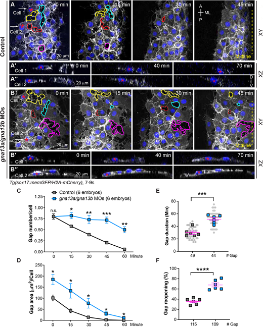
Fig. 3 Gα13 regulates stable cell-cell contacts and proper changes of endodermal cell shape during endoderm C&E. Confocal time-lapse experiments were conducted at the most lateral and anterior regions of pharyngeal endoderm in the indicated embryos during 7-9 ss. (A,B) Snapshots of confocal z projections in the xy view, taken at different time points from Movie 1, illustrating the morphology of endodermal cells at various time points. Gaps between endodermal cells are outlined, and the same gap over time is outlined by the same color. (A'-B″) Confocal images of xz planes of two representative cells (cell 1 and cell 2), captured at regions marked by white arrows in A and B, from Movie 2, illustrating the orientation of the nuclei of two cells at the indicated time-points. (C-F) The average number (C), area (D), duration (E) and reopening frequency (F) of the gaps in endodermal cells in the indicated embryos at different time points. (E) Data from all embryos (squares) and all xy images (gray circles) are superimposed. A, anterior; P, posterior; ML, mediolateral; D, dorsal; V, ventral. Yellow dashed line, midline. Data are mean±s.e.m. n.s. (not significant), P>0.05; *P<0.05; **P<0.01; ***P<0.001; ****P<0.0001 (unpaired, two-tailed Student's t-test).