
Fig. S2
- ID
- ZDB-IMAGE-241009-9
- Publication
- Ugur et al., 2024 - VPS13B is localized at the interface between Golgi cisternae and is a functional partner of FAM177A1
- All Figures
- Figures for Ugur et al., 2024
|
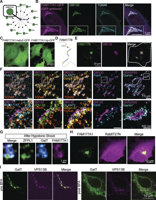
Fig. S2 Human FAM177A1 and FAM177B localize at the Golgi complex. (A) Bioplex data showing predicted partners of VPS13B (https://bioplex.hms.harvard.edu/explorer/network.php). (B) HeLa cells expressing FAM177A1-GFP immunolabeled with anti-GFP, anti-GM130, and anti-TGN46 antibodies. (C) COS7 cells expressing FAM177A1-helix2-GFP (left) or FAM177A1-Hairpin-GFP (right). (D) Alphafold2 predicted structure of FAM177B. (E) HeLa cells expressing FAM177B-flag fixed and immunolabeled with anti-flag and anti-GM130 antibodies. n: nucleus, scale bar = 10 µm. (F) FLASH-PAINT performed in HeLa cells expressing VPS13B^GFP and FAM177A1-Halo and immunolabeled with antibodies directed against GFP, halo, GM130, GLANT2, Golgin-97, Rab6, TGN46, GRASP65, and Giantin. (G) Snapshots of the Golgi complex of a HeLa cells expressing ZFPL1-GFP, GalT-RFP, and FAM177A1^Halo after a 10-min hypotonic shock. Scale bar = 1 µm. (H) HeLa cells expressing FAM177A1-GFP and Rab6T27N-RFP. Scale bar = 10 µm. (I) HeLa cells expressing GalT-RFP and VPS13B^Halo before (left panel) and after BFA treatment (5 µg/ml for 40 min, right panel). Scale bar = 10 µm.