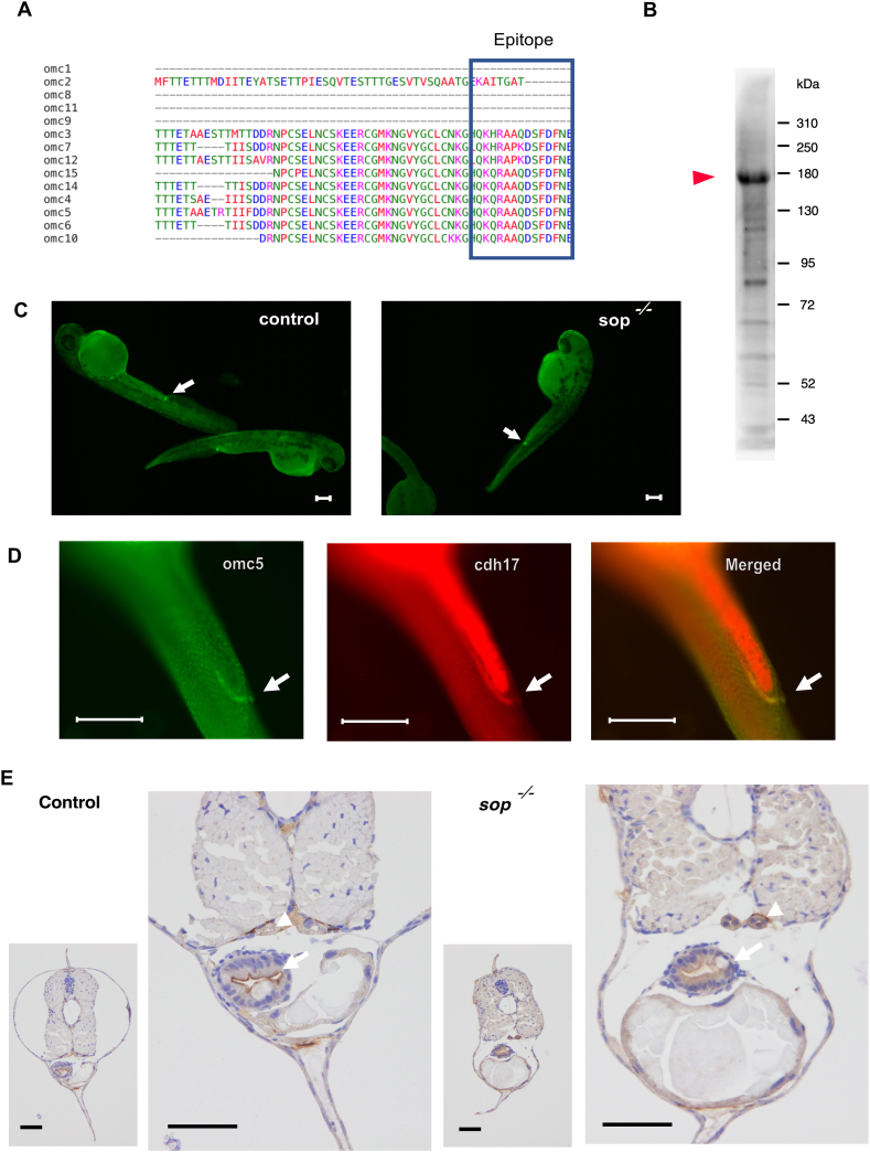
IMAGE

Fig. 2

ID
ZDB-IMAGE-240923-4
Source
Figures for Naruoka et al., 2024
Image
Figure Caption

Fig. 2

A. Amino acid sequence of omcin gene products surrounding the epitope used for the generation of antibody (boxed in blue). 6 of omcin genes including omc5 have the identical sequence for the epitope, while additional 3 genes have close sequences. B. Western blot of the Omcin antibody on5dpf embryos. The strongest signal was observed around 180 kDa (arrowhead).C. Antibody signals observed near cloaca in control and sop−/− embryos (arrows).D. Double staining in wild type embryos of Omcin antibody and cdh17. Signals indicated by arrows overlapped in the distal pronephros.E. Omcin antibody staining in slices of control and sop−/−embryos at 5 dpf. Signals in the gut (arrows) and the pronephros (arrowheads) are indicated. Scale bars, 50 μm.

Acknowledgments
This image is the copyrighted work of the attributed author or publisher, and ZFIN has permission only to display this image to its users. Additional permissions should be obtained from the applicable author or publisher of the image. Full text @ Heliyon