
|
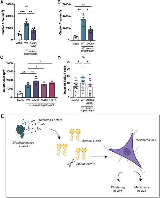
Fig. 5 Deletion of S. aureus lipases alters melanoma clustering and invasion. Quantification of ZMEL1 melanoma cell cluster size (µm2) after 7 days of culture in RPMI medium alone or plus S. aureus USA300 bacterial supernatant as indicated. (A–C) Supernatants were tested from USA300 transposon mutants for lipases Sal2/Geh (Δ0320) (A), phosphatidylinositol-specific phospholipase C (Δ0099) (B) or for putative phospholipase genes Δ2457, Δ0070 and Δ1710 (C). PRMI medium alone (Media) was used as control. (D) ZMEL1 invasion at 2 dpi in the zebrafish hindbrain after culture in medium alone or in S. aureus USA300 supernatant. Dots represent independent larvae color coded per replicate. Medium control (n=30 larve); WT bacterial supernatant (n=32 larvae); Δ0320 supernatant (n=24 larvae). Experiments were conducted at least three times. Dots in A-C represent independent replicates. Error bars indicate the mean±s.e.m. P-values were calculated by one-way ANOVA (A–D). *P<0.05; **P<0.01; ***P<0.001. ns, not significant. (E) Schematic summarizing the findings described in this article, showing that S. aureus secreted factors promote melanoma clustering in vitro and invasion in vivo.