
|
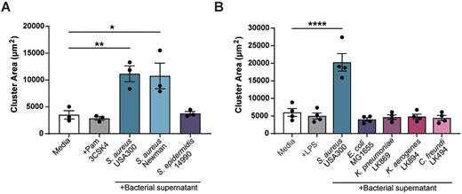
Fig. 3 Melanoma clustering is specific to S. aureus species. Quantification of ZMEL1 melanoma cell cluster size (µm2) after 7 days of culture in either RPMI medium alone (Media) or in RMPI medium plus bacterial supernatant. (A) RPMI medium alone (Media), or with addition of Pam3CSK4 (100 µg/ml) or with one of the bacterial supernatants obtained from a selection of cultured Gram-positive bacteria as indicated was added to melanoma cell culture. (B) RPMI medium alone (Media), Media plus LPS (1 µg/ml) or with one of the bacterial supernatants obtained from a selection of cultured Gram-negative bacteria as indicated was added to melanoma cell culture. Experiments were conducted at least three times. Dots represent independent replicates. Error bars indicate the mean±s.e.m. P-values were calculated by one-way ANOVA. *P<0.05; **P<0.01; ****P<0.0001.