
Image
|
Figure Caption
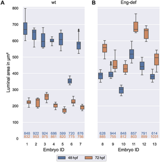
Fig. 9 High between-embryo but low within-embryo variability in luminal area. Distribution of luminal areas of estimated DA cross-sections, stratified by embryo and time, for wild-type embryos (A) and for Endoglin-deficient embryos (B). Analysis based on cross-sections with at least two annotated cells present within a distance of 0.5 μm to the cross-sectional plane. The numbers below the box plots denote the corresponding numbers of vessel cross-sections. The top number corresponds to 48 hpf and the bottom number to 72 hpf.
Acknowledgments
This image is the copyrighted work of the attributed author or publisher, and
ZFIN has permission only to display this image to its users.
Additional permissions should be obtained from the applicable author or publisher of the image.
Full text @ PLoS Comput. Biol.