
Fig. 6
- ID
- ZDB-IMAGE-240909-16
- Publication
- Campbell et al., 2024 - p65 signaling dynamics drive the developmental progression of hematopoietic stem and progenitor cells through cell cycle regulation
- All Figures
- Figures for Campbell et al., 2024
|
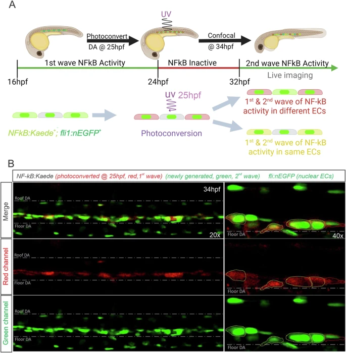
Fig. 6 In vivo NF-kB signaling dynamics at the single cell resolution.A Illustration depicting the experimental design in (B). Briefly, NFkB:Kaede+; fli:nEGFP+ embryos were photoconverted at 25hpf with UV light for 10 min and subsequently subjected to confocal microscopy in the DA at 34hpf. B Representative maximum projection confocal images of the DA of 34hpf NFkB:Kaede+; fli:nEGFP+ embryos. Yellow (Kaede green+; Kaede red+) cytoplasms are cells with both NF-kB activity waves occurring within the same cell (cells outlined in yellow). Dashed red delineates EC with Kaede (red+, green-) cytoplasm. The experiment was repeated three times independently with similar results. Figure 6A created with BioRender.com released under a Creative Commons Attribution-NonCommercial-NoDerivs 4.0 International license.