
Fig. 5
- ID
- ZDB-IMAGE-240909-15
- Publication
- Campbell et al., 2024 - p65 signaling dynamics drive the developmental progression of hematopoietic stem and progenitor cells through cell cycle regulation
- All Figures
- Figures for Campbell et al., 2024
|
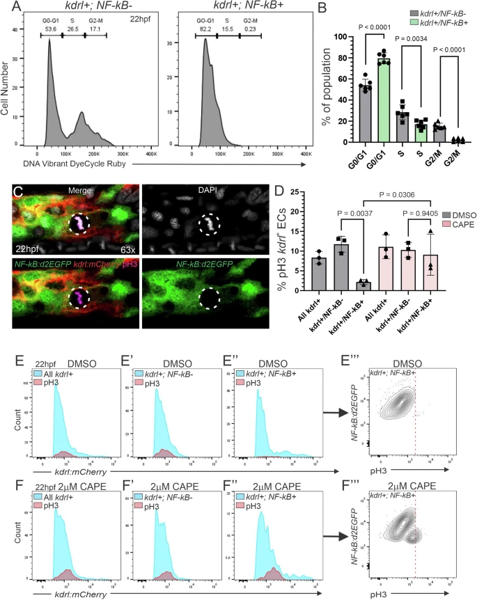
Fig. 5 P65 drives EC quiescence prior to endothelial to hematopoietic transition.A Representative histograms of live kdrl:mCherry+; NF-kB:d2EGFP− (left) and kdrl:mCherry+; NF-kB:d2EGFP+ cells (right) from 15–20 pooled 22hpf isolated trunks stained with DNA Vibrant DyeCycle Ruby. Brackets denote resting/Gap 1 phase (G0-G1), Synthesis phase (S), and Gap 2/Mitotic phase (G2-M). B Quantification from (A). Each dot represents 15–20 pooled embryonic trunks, n = 6. Black horizontal lines indicate mean ± SD. C Representative confocal maximum projection image of the DA of a 22hpf NF-kB:d2EGFP+; kdrl:mCherry+ embryo subjected to WIHC for pH3 (magenta), NF-kB activation (green), and the endothelial marker kdrl (red). The dashed circle indicates a mitotic pH3+ cell. Notice that mitotic kdrl+ ECs have inactive NF-kB (NF-kB−). The experiment was repeated three times independently with similar results. D Quantification of the percentage of pH3+ cells from (E, E”, F, F”). Each dot represents 15–20 pooled embryonic trunks, n = 3. Black horizontal lines indicate mean ± SD. (E, F”’) Representative histograms depicting flow cytometric analysis of 22hpf DMSO (E, E”’), or 2 µM CAPE-treated (F, F”’), dissociated NF-kB:d2EGFP+; kdrl:mCherry+ embryonic trunks subjected to intracellular flow for d2EGFP, mCherry (blue), and pH3 (red). Chemical treatments were applied from 16hpf. E, F All kdrl:mCherry+ cells. E’, F’ kdrl:mCherry+; NF-kB:d2EGFP− cells. E”, F” kdrl:mCherry+; NF-kB:d2EGFP+ cells. E”’, F”’ Representative contour plot of kdrl:mCherry+; NF-kB:d2EGFP+ cells from (E”). The dotted line separates pH3− (left) from pH3+ (right) cells. Data were analyzed by ordinary one-way ANOVA with Turkey’s multiple comparisons test (B, D). Source data are provided as a Source Data file.