
IMAGE
Fig. 1
- ID
- ZDB-IMAGE-240904-6
- Publication
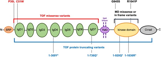
- Monaghan et al., 2024 - FLT4 causes developmental disorders of the cardiovascular and lymphovascular systems via pleiotropic molecular mechanisms
- All Figures
- Figures for Monaghan et al., 2024
Image
|
Figure Caption
Fig. 1 Schematic representation of FLT4/VEGFR3 protein. Red bracket: location of missense or in frame indels identified in TOF. Black bracket: location of inframe MD variants. Blue bracket: location of TOF-PTVs (P30fs*3 to Y1337fs*19). The eight variants experimentally studied here are indicated. SRP, signal recognition peptide; IgDs, immunoglobulin-like domains.
Acknowledgments
This image is the copyrighted work of the attributed author or publisher, and
ZFIN has permission only to display this image to its users.
Additional permissions should be obtained from the applicable author or publisher of the image.
Full text @ Cardiovasc. Res.