
|
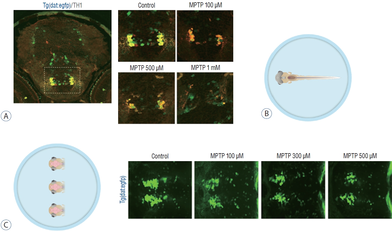
Fig. 2 Zebrafish preparation for quantification of dopaminergic neurons. A : Immunostaining for tyrosine hydroxylase (TH) on horizontal cryosection of Tg(dat:EGFP) larvae at 3 days post-fertilization (dpf) following 1-methyl-4-phenyl-1,2,3,6-tetrahydropyridine (MPTP) treatment for 48 hours from 1 dpf with each MPTP concentration. B : In vivo imaging technique by fixing the whole larva ventrally. C : In vivo imaging technique by fixing the heads of larvae. Representative morphology of dopaminergic neurons in Tg(dat:EGFP) by in vivo imaging with each MPTP concentration. Ventral view, head at the left, tectum at the right.