
|
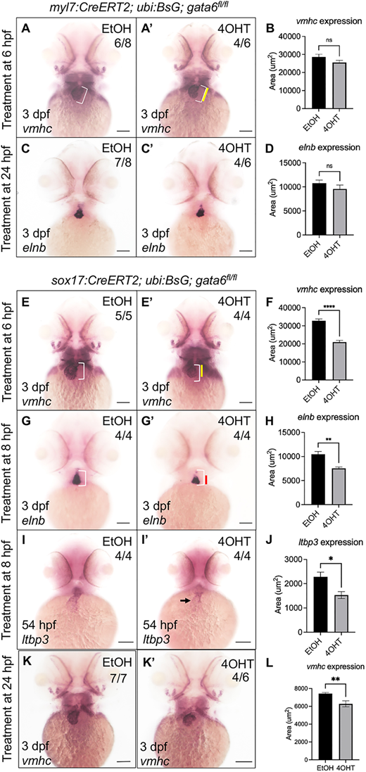
Fig. 7 Loss of endodermal but not myocardial gata6 expression disrupts development of the anterior secondary heart field. (A-D) Loss of myocardial gata6 does not significantly impair cardiac development. Whole-mount in situ hybridization (WISH) staining showed no significant difference in vmhc (A,A′) or elnb (C,C′) expression domains in gata6fl/fl embryos after ethanol or tamoxifen treatment at 6 hpf or 24 hpf, respectively. White brackets indicate the length of the ventricle in ethanol-treated controls. Yellow bar indicates the length of the ventricle in tamoxifen-treated embryos. (B,D) Quantification of the area of WISH staining (paired t-test; ns, not significant). Data are mean±s.e.m. (E-H) Loss of endodermal gata6 disrupts cardiac morphogenesis. (E,E′) WISH analysis for vmhc expression pattern in gata6fl/fl embryos revealed a notable reduction in the size of the ventricle after tamoxifen treatment at 6 hpf. White brackets indicate the length of the ventricle in ethanol-treated controls. Yellow bar indicates length of ventricle in tamoxifen-treated embryos. (G,G′) WISH analysis for elnb transcript patterns revealed shortened outflow tracts in gata6fl/fl hearts after tamoxifen treatment at 8 hpf. White brackets indicate the length of the outflow tract in ethanol-treated controls. Red bar indicates the length of the outflow tract in tamoxifen-treated embryos. (F,H) Quantification of the area of WISH staining confirmed a significant loss of expression domain within the heart (F) and outflow tract (H) (paired t-test, ****P<0.0001 and **P<0.005, respectively). Data are mean±s.e.m. (I,I′) Loss of endodermal gata6 reduces ltbp3 expression levels. WISH analysis revealed decreased expression domain of ltbp3 transcripts in tamoxifen-treated gata6fl/fl embryos (I′) compared with ethanol-treated controls (I). Black arrow indicates reduced expression of ltbp3 within the ventricle. (J) Quantification of WISH staining showed a significant reduction in the area of ltbp3 mRNA expression within the heart (paired t-test, *P<0.05). Embryos were treated overnight starting at 8 hpf. (K-L) Late removal of endodermal gata6 impairs ventricular chamber development. Representative WISH images of 3 dpf Tg(sox17:CreERT2); Tg(ubb:BsG); gata6fl/fl embryos after exposure to ethanol (K) or 10 μM tamoxifen (K′) from 24 hpf to 48 hpf analyzed with probes targeting vmhc. (L) Quantification of the area of vmhc expression showed a significant reduction in tamoxifen-treated embryos compared with control (paired t-test, **P<0.01, data are mean±s.e.m.). Scale bars: 100 µm. Area of staining was measured manually using FIJI software.