
|
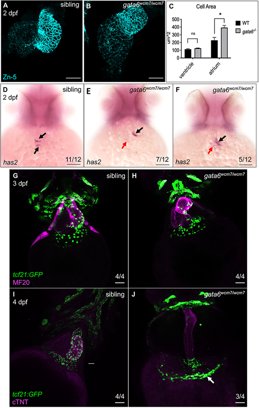
Fig. 1 Loss of gata6 impairs heart morphology. (A-C) Atrial cardiomyocytes (CMs) are enlarged in gata6wcm7/wcm7 hearts. (A) Immunostaining of dissected wild-type (A) or gata6 mutant (B) hearts showing expression of the cell membrane marker Zn-5. The size of the atrial chamber in gata6 mutants is noticeably extended compared with wild type. Scale bars: 50 µm. (C) Quantification of cell area showed a significant increase in atrial but not ventricular CM cell area in gata6 mutant embryos (paired t-test, *P<0.05). The area of 10 random cells from each chamber was quantified from three different hearts for each genotype. Data are mean±s.e.m. (D-F) Loss of gata6 impairs atrioventricular canal (AVC) development. Whole-mount in situ hybridization staining revealed decreased expression of has2 transcripts in gata6 mutant hearts (E,F) compared with siblings (D). Arrows indicate normal (black) or much reduced (red) mRNA expression level in the AVC. Scale bars: 100 µm. (G-J) gata6wcm7/wcm7 epicardial cells fail to migrate across the myocardium. Immunostaining indicated successful migration of tcf21:GFP+ epicardial cells at the base of the ventricular myocardium of sibling (G) and gata6 mutant (H) hearts at 3 dpf. Subsequently, at 4 dpf, imaging revealed tcf21:GFP+ cells covering the sibling ventricles (I) but clustering mostly near the venous pole of gata6wcm7/wcm7 hearts (J). White arrow indicates the area of cell accumulation. Scale bars: 50 µm.