
|
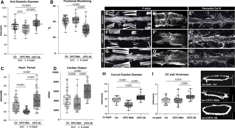
Fig. 2 The effects of CRTC are cardiac autonomous (A) Cardiac-specific CRTC KD with tinCΔ4-Gal4 significantly reduced EDD, while cardiac CRTC OE increased EDD, compared to controls (tinCΔ4-Ga4/+). (B) Fractional shortening was significantly reduced by cardiac CRTC OE. (C) Heart period was significantly reduced in CRTC-KD hearts and increased in hearts with CRTC OE. (D) Cardiac output was reduced in CRTC-KD hearts and increased in hearts with CRTC OE. All flies were 3 weeks of age; plots show all data points, max, min, median, and p values; significance by one-way ANOVA with Tukey’s multiple comparisons post hoc test. (E–G) (E) F-actin staining shows sarcomeric structure in cardiac tubes and adjacent body wall muscles. (E′) Optical section through one cardiac chamber showing the tightly packed, circumferential fibers (arrowhead) in a control heart. (Asterisk denotes position of the ostia, anterior is left in all pictures.) (F) F-actin staining of a heart with cardiac CRTC KD, exhibiting cardiac restriction and (F′) disorganized myofibrils with malformed ostia. (G) Cardiac CRTC-OE heart showing cardiac dilation and (G′) some disorganized myofibrils and gaps and malformed ostia. (E″) Wild-type control stained for collagen IV (pericardin) reveals the extensive extracellular matrix. (F″) The collagen matrix in CRTC KD is expanded, especially in the posterior heart (right), but (G″) not with CRTC OE. (For E–G″, scale bars represent 40 μm). (H) Diameters from optical sections of dystroglycan-stained hearts. Cardiac conical chamber (CC) diameters were significantly reduced with cardiac CRTC KD and increased with cardiac CRTC OE. (I) Cardiomyocyte thickness, measured from optical sections of dystroglycan-stained hearts (to the right, scale bar represents 50 μm), was increased with cardiac CRTC OE compared to control and cardiac CRTC-KD hearts.