
|
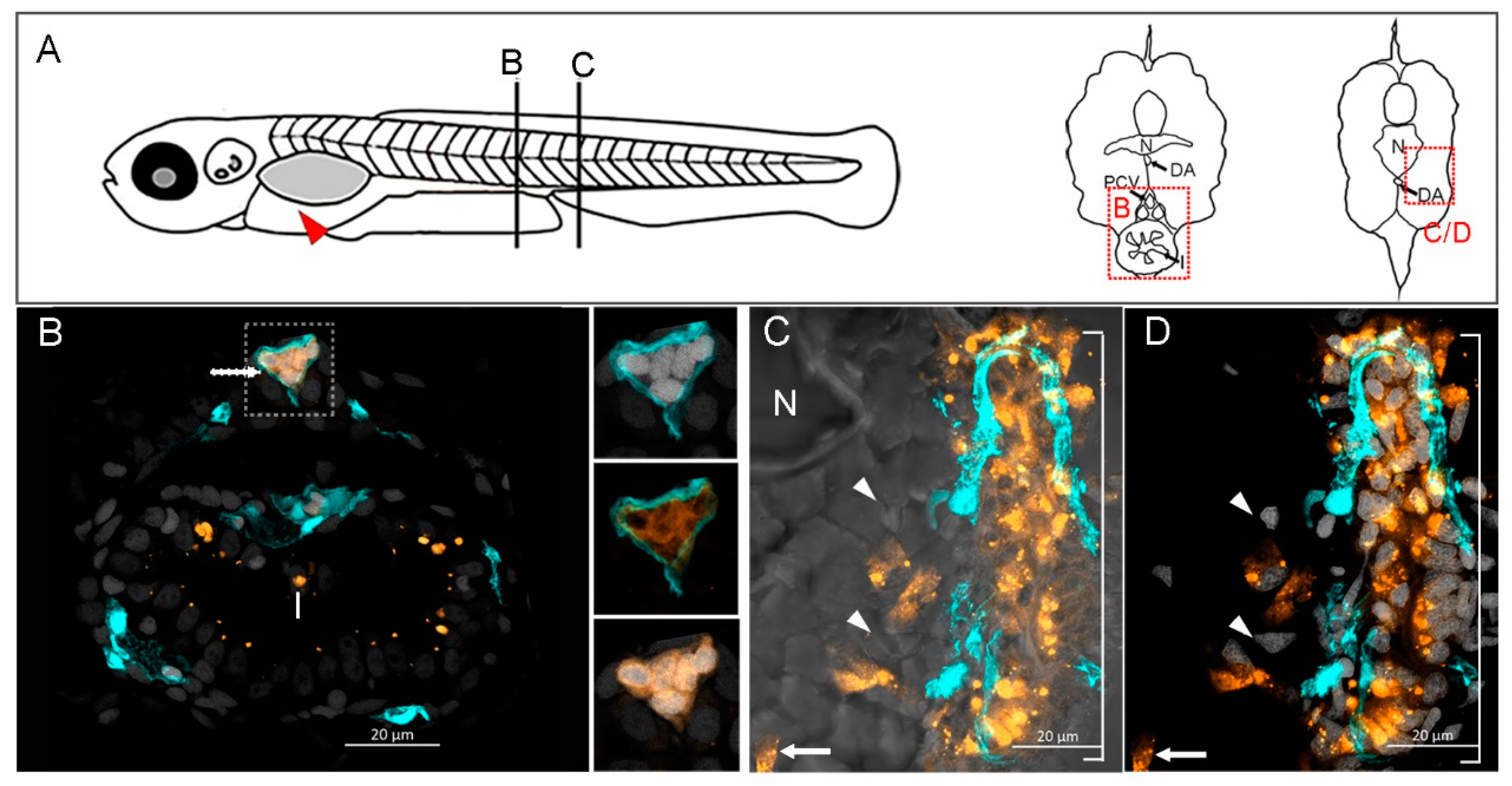
Fig. 7 GCSCs migrate in groups and establish metastatic tumors. (A) Tg (fli1:EGFP)y1 zebrafish embryos (2 dpf) were given an injection into the yolk sac (red arrow) of 50 fluorescently observed migrating cells (white arrow) inside the posterior cardinal vein (PCV, cyan) over the distal portion of the intestine (I). The insets show five GCSC nuclei inside the PCV, co-stained with Hoechst (white). (C,D) The GCSCs formed a metastatic cell mass that invaded the skeletal muscle at the level of somites 21-21, as observed using a brightfield microscope (C), the nuclei were stained with Hoescht (D) to observe clearly the cell mass. The white arrows show GCSCs, and the white arrowheads indicate zebrafish muscle fiber nuclei. N indicates the notochord, DA the dorsal aorta, and I the intestine. The images were obtained at 63X with an LSM800 confocal microscope. Labeled GCSCs are shown in orange, and 6 µm sections are shown in the (B,C) insets. (B) GCSCs 6 dpi.