
|
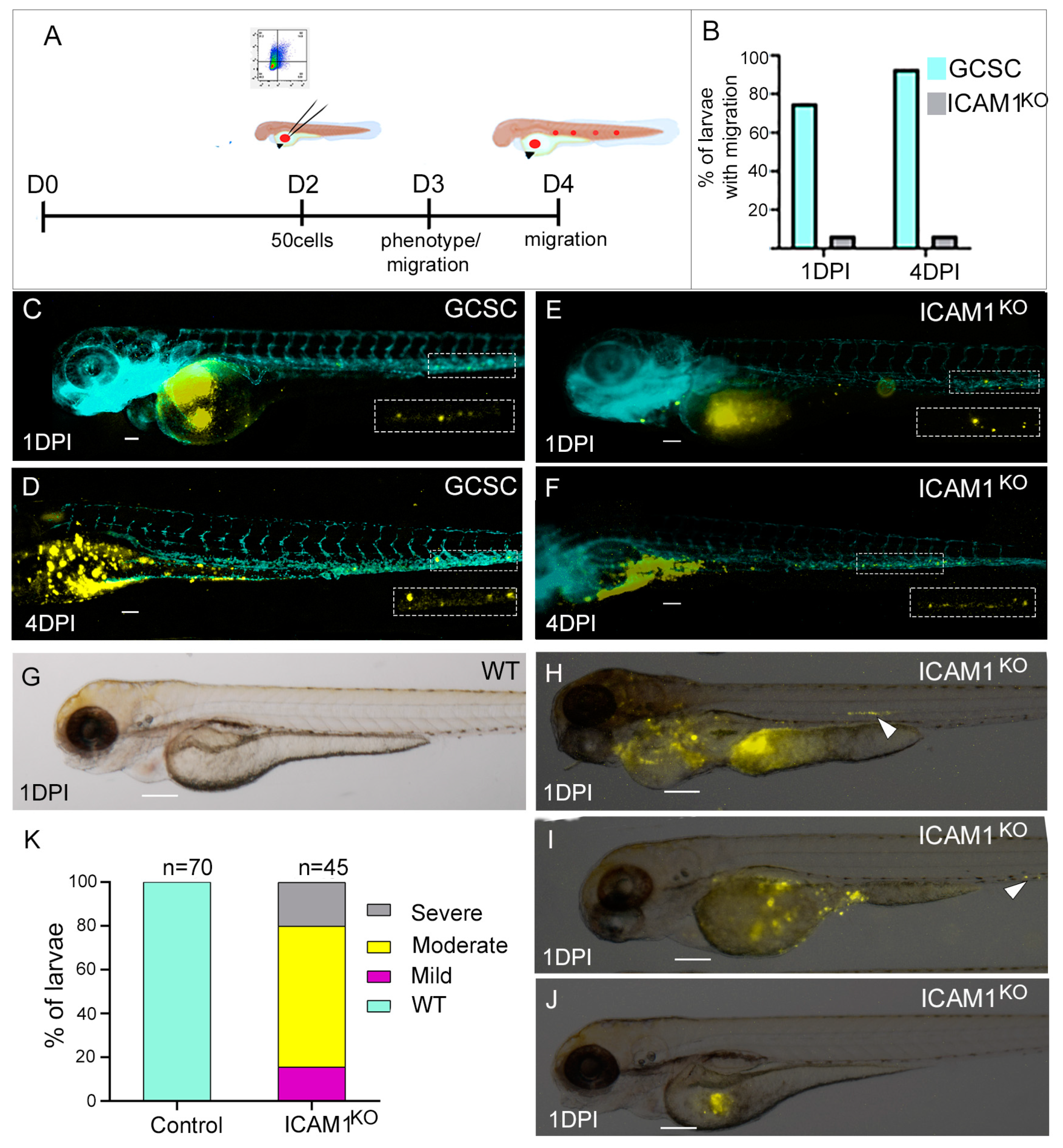
Fig. 6 The tumorigenic and migratory capabilities of xenotransplanted GCSC/ICAM1KO cells. (A) The experimental design. (B) The percentage of embryos with migrating GCSCs or GCSC/ICAM1KO cells after 1 and 4 dpi. (C,D) show larvae injected with GCSCs after 1 or 4 dpi, respectively. (E,F) show larvae injected with GCSC/ICAM1KO cells after 1 or 4 dpi, respectively. (H–J) show larvae after 1 dpi with severe, moderate, or mild phenotypes, respectively. (G) shows a wild-type larva. (K) shows the percentage of larvae with severe, moderate, or mild phenotypes (n = 45). The images were obtained with a Nikon SMZ1500 stereomicroscope. All the bars indicate 100 µm. GCSC = AGS/GCSC, and ICAM1KO = GCSC/ICAM1KO. All boxes and arrow heads indicate sites with migrating cells.