
|
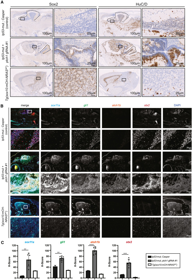
Fig. 5 Zebrafish ptch1-crispant brain tumors are heterogeneous for neural stem cells and differentiated neurons with elevated SHH-pathway genes (A) Immunohistochemistry of Sox2 (left) and HuC/D (right) in the indicated genetic backgrounds. A higher magnification of the black-boxed area is shown in the adjacent panels to the right. Black dashed lines indicate the cerebellum. (B) Immunofluorescent RNAscope in the indicated genetic backgrounds with probes against sox11a, gli1, atoh1b, and otx2. Individual probes are shown in grayscale. White boxes in the upper left are shown at higher magnification below. DAPI was used as a nuclear marker. (C) The H score was independently calculated for each RNAscope probe and is plotted with the standard deviation. Each dot represents an individual section. Statistical significance was calculated using an unpaired two-tailed t test. ∗∗p < 0.006, ∗∗∗p = 0.0004, ∗∗∗∗p < 0.0001. See also Figures S1 and S2.