
|
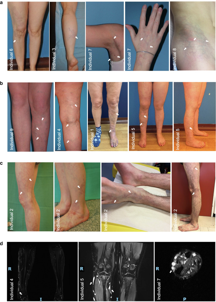
Fig. 2 Clinical phenotype of patients with CMDV with PIK3R1 or nonhot post-PIK3CA variants. Patients characterized by localized CMs and dilated veins visible on skin and MRI (white arrows). (a) Patients with smaller lesions and less prominent veins. (b) Patients with more extensive lesions and strongly prominent veins. (c) Progression of lesion in individual 2 from the age 7 to the age of 21 year: veins became more prominent and dilated, and CM turned from reddish to purplish color. (d) MRI (fat suppression sequence, 1.5T) of the lower limbs of patients 4, 5, and 7 showing hyperintense slow-flowing blood in dilated veins of the subcutaneous tissues. All subjects or their parent/guardian included in this study consented to publication of the images shown in this figure. CM, capillary malformation; CMDV, capillary malformation with dilated veins; MRI, magnetic resonance imaging.