
|
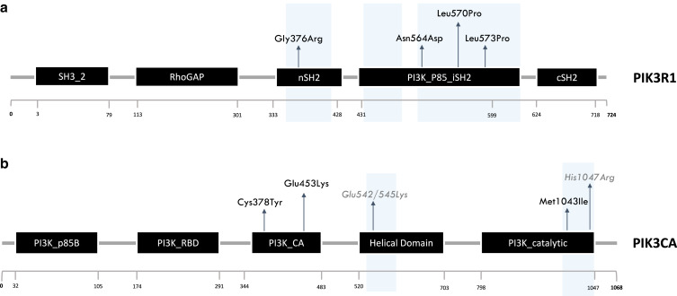
Fig. 1 PIK3CA and PIK3R1 protein structures and distribution of identified variants. (a) PIK3R1 protein (p85) and its functional domains. Variants localized in the nSH2 domain (n = 1) and in the PI3K_P85_iSH2 domain (n = 3). L570P and L573P variants are predicted to be pathogenic but lack experimental validation. (b) PIK3CA (p110) and its functional domains. Variants reported in black localized in the Ca2+-binding domain (n = 2) and in PI3K catalytic domain (n = 1). Hotspot PIK3CA mutations are in grey. Cancer hotspot regions are highlighted in light blue. Ca2+, calcium ion; PI3K, phosphoinositide 3-kinase.