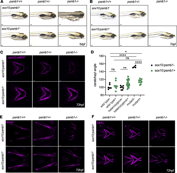
IMAGE

Figure 6

ID
ZDB-IMAGE-240826-8
Source
Figures for Miller et al., 2024
Image
Figure Caption

Figure 6 Overexpression of psmb1 in the neural crest and chondrocytes rescues mutant cartilage and tendon phenotype.

(A) Bright-field images of psmb1 wild-type, heterozygous, and mutant larvae with and without sox10:psmb1-2A-GFP transgene at 120 hpf. Scale bars: 100 μm. (B) Bright-field images of psmb1 wild-type, heterozygous, and mutant larvae with and without sox10:psmb1-2A-GFP transgene at 7 dpf. Scale bars: 100 μm. (C) Confocal imaging of sox10:mRFP, psmb1 wild-type, heterozygous, and mutant larvae with and without sox10:psmb1-2A-GFP transgene at 72 hpf. Scale bars: 50 μm. (D) Quantification of ceratohyal angle in images from C demonstrates that ceratohyal angle is almost completely restored in rescued psmb1 mutants. Data shown represent mean ± SD. n = 6, 8, 7, 22, 4, 10. Significance was calculated with 2-way ANOVA. *P < 0.05, **P < 0.01, ****P < 0.0001; ns, not significant. (E) sox10:psmb1-2A-GFP transgene substantially improves tendon phenotype, as assessed by RNAscope for tnmd at 72 hpf. Scale bars: 50 μm. (F) sox10:psmb1-2A-GFP partially rescues muscle phenotype, as assessed by antibody staining for myosin heavy chain (MHC) at 72 hpf. Scale bars: 50 μm.

Figure Data
Acknowledgments
This image is the copyrighted work of the attributed author or publisher, and ZFIN has permission only to display this image to its users. Additional permissions should be obtained from the applicable author or publisher of the image. Full text @ JCI Insight