|
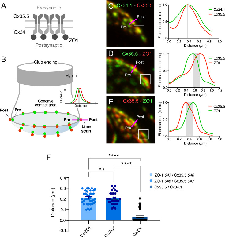
Figure 4. Expansion microscopy reveals the molecular components of gap junction (GJ) plaques at club ending (CE) synaptic contact areas.
(

|
|
Figure 4. Expansion microscopy reveals the molecular components of gap junction (GJ) plaques at club ending (CE) synaptic contact areas.
(