
|
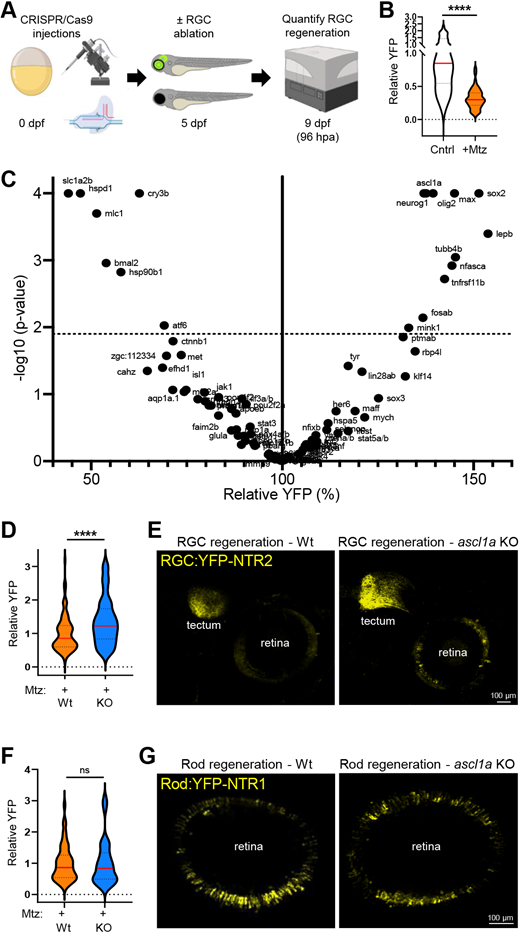
Fig. 4 CRISPR screen identifies genes regulating RGC regeneration. (A) Experimental setup for CRISPR/Cas9-based ‘crispant’ knockout (KO) screen for genes that regulate RGC regeneration. (B) Plate reader-based quantification of RGC regeneration at 96 hpa. Relative YFP levels in RGC ablated larvae (+Mtz) are normalized to unablated controls (Cntr). (C) Volcano plot of RGC regeneration (relative YFP levels) for all Mtz-treated RGC:YFP-NTR2 crispant KO larvae (dashed line indicates adjusted P-value cutoff of 0.01; minimum sample size n=8, median sample size n=16). (D) Plate reader-based quantitative comparison of RGC regeneration levels (YFP) between Mtz-treated control (Wt) and ascl1a crispant (KO) larvae at 96 hpa. (E) Representative confocal images of RGC regeneration in Mtz-treated control (Wt) and ascl1a crispant (KO) larvae at 168 hpa. (F) Plate reader-based quantitative comparison of rod cell regeneration levels (YFP) between Mtz-treated control (Wt) and ascl1a crispant (KO) larvae at 96 hpa. (G) Representative confocal images of rod cell regeneration in Mtz-treated control (Wt) and ascl1a crispant (KO) larvae at 168 hpa. ****P≤0.0001; ns, not statistically significant.