
|
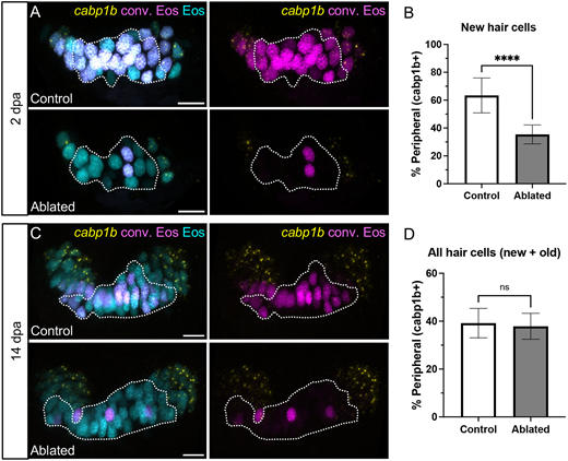
Fig. 7 Hair cell central-peripheral patterning is restored after ablation. (A) Representative maximum intensity projections of anterior crista in control and ablated fish at 2 dpa with cabp1b HCR-FISH. Photoconverted Eos (magenta) and cabp1b (yellow) channels are shown with and without unconverted Eos (cyan). Dotted outline delineates the central cabp1b− region of the sensory patch. (B) Quantification of cabp1b+ new hair cells, shown as a percentage of all new (cyan only) hair cells in control (n=18) and ablated (n=16) anterior cristae. Unpaired t-test, ****P<0.0001. (C,D) Analogous data to A,B for crista at 14 dpa (n=18 control, 15 ablated). Unpaired t-test, P=0.5226. Scale bars: 10 µm. Data are mean±s.d.