
|
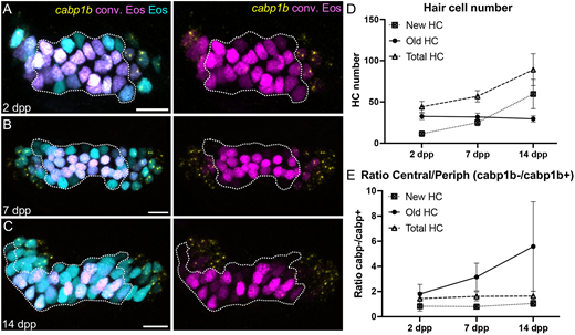
Fig. 4 Identification of inner ear hair cell subtypes during larval growth. (A-C) Maximum intensity projection images of HCR-FISH probing for cabp1b expression in Tg(myo6b:NLS-Eos) anterior cristae at (A) 2 days post-photoconversion (dpp) (10 dpf, n=14); (B) 7 days dpp (15 dpf, n=12); and (C) 14 dpp (22 dpf, n=8). Old hair cells retain photoconverted (magenta) Eos signal while new hair cells have unconverted (cyan) Eos only. Peripheral-type hair cells are labeled by the cabp1b HCR probe (yellow). Dotted outline delineates the central cabp1b− region of the sensory patch. Scale bars: 10 µm. (D) Increase in hair cell numbers over the course of the experiment. (E) Ratio of central (cabp1−) to peripheral (cabp1b+) hair cells over time. The increased ratio for old cells suggests phenotypic conversion from peripheral to central hair cell type over time. Data are mean±s.d.