
|
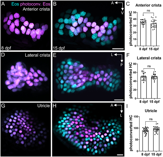
Fig. 3 Little hair cell turnover occurs in the larval zebrafish ear. (A,B) Representative maximum intensity projection images of Tg(myo6b:NLS-Eos) anterior cristae (A) immediately post-photoconversion at 8 days post-fertilization (dpf) or (B) 1 week post-photoconversion at 15 dpf. Hair cells that were photoconverted retained photoconverted (magenta) Eos signal, while new hair cells have unconverted (cyan) Eos only. (C) Quantification of anterior crista photoconverted hair cells at 8 and 15 dpf (n=20 at 8 dpf, 20 at 15 dpf). (D-I) Analogous results for the lateral crista (n=20, 20) (D-F) and for the utricle (n=29, 25) (G-I). Unpaired t-tests indicate no significant difference between the number of photoconverted hair cells at these two timepoints (anterior crista P=0.125, lateral crista P=0.859, utricle P=0.071). Scale bars: 10 µm. Data are mean±s.d.