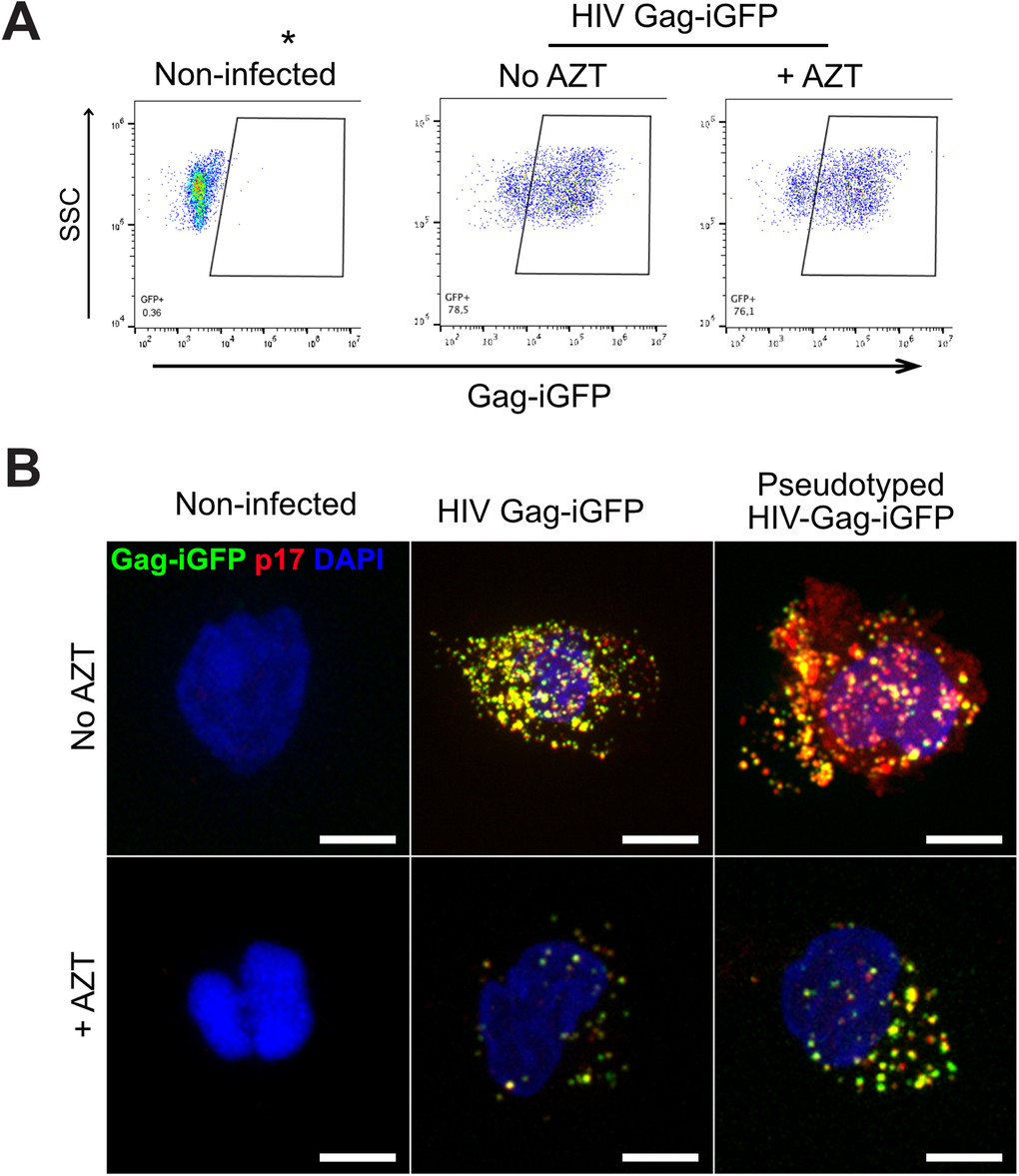
Fig. EV5 Characterization of primary monocyte infection by HIV-1 and transmigration. (A) Primary monocytes were non-infected or infected with HIV-1 (NLAD8) at MOI 1 for 48 h in the presence or absence of 10 µM AZT. The dot plots show the percentage of Gag-iGFP-expressing cells as a function of the side scatter (SSC) measurement as determined by flow cytometry. The data highlights that despite AZT treatment, monocytes are positive for Gag-iGFP as they carry fluorescent particles. (B) Primary monocytes were non-infected, infected with HIV-1 Gag-iGFP, or HIV-1 Gag-iGFP pseudotyped with VSV-G, at MOI 1 for 48 h in the presence or absence of AZT. Cells were fixed and stained for Gag p17 (red) and DAPI (blue) and Gag-iGFP is shown in green. Scale bar: 5 µm.
Image
Figure Caption
Acknowledgments
This image is the copyrighted work of the attributed author or publisher, and
ZFIN has permission only to display this image to its users.
Additional permissions should be obtained from the applicable author or publisher of the image.
Full text @ EMBO Rep.

