
Fig. 7
- ID
- ZDB-IMAGE-240808-31
- Publication
- Xia et al., 2024 - A Novel Role for the Longevity-Associated Protein SLC39A11 as a Manganese Transporter
- All Figures
- Figures for Xia et al., 2024
|
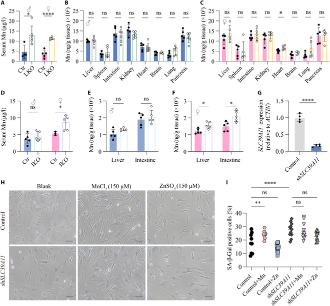
Fig. 7 Tissue-specific Slc39a11 knockout mice have increased serum Mn levels, and knocking down SLC39A11 in vitro increases cellular senescence. (A to C) Summary of Mn levels measured in the serum (A) and indicated tissues (B and C) in 2-month-old Slc39a11flox/flox (Ctr) and liver-specific Slc39a11 knockout (LKO) male (B) and female (C) mice. (D to F) Summary of Mn levels measured in the serum (D) and liver and intestine (E and F) of 2-month-old Slc39a11flox/flox (Ctr) and intestine-specific Slc39a11 knockout (IKO) male (E) and female (F) mice. (G) Summary of SLC39A11 mRNA measured in control fibroblasts and fibroblasts expressing an shRNA against SLC39A11 (shSLC39A11). (H) Representative phase contrast images of control fibroblasts and fibroblasts expressing shSLC39A11 stained for SA-β-Gal after exposure to no additional metal ions (blank), 150 μm MnCl2, or 150 μM ZnSO4. Scar bar, 10 µm. (I) Summary of the percentage of SA-β-Gal-positive cells treated as indicated. *P < 0.05, **P < 0.01, ****P < 0.0001.