
Fig. 4
- ID
- ZDB-IMAGE-240808-28
- Publication
- Xia et al., 2024 - A Novel Role for the Longevity-Associated Protein SLC39A11 as a Manganese Transporter
- All Figures
- Figures for Xia et al., 2024
|
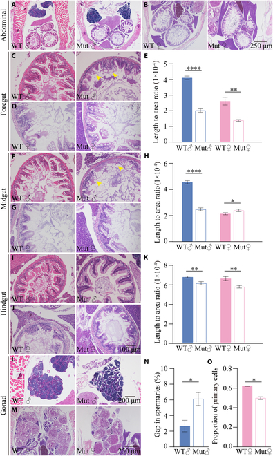
Fig. 4 slc39a11 Mut zebrafish have altered visceral organ and gonad morphology. (A and B) H&E-stained abdominal sections prepared from male (A) and female (B) WT and Mut zebrafish. (C to E) H&E-stained foregut sections prepared from male (C) and female (D) WT and Mut zebrafish, and summary of the ratio between villus length and foregut area (E). (F to H) H&E-stained midgut sections prepared from male (F) and female (G) WT and Mut zebrafish, and summary of the ratio between villus length and midgut area (H). (I to K) H&E-stained foregut sections prepared from male (I) and female (J) WT and Mut zebrafish, and summary of the ratio between villus length and foregut area (K). (L and M) H&E-stained spermary (L) and ovary (M) sections prepared from male (L) and female (M) WT and Mut zebrafish. (N and O) Summary of the percentage of gaps in the spermaries of male WT and Mut zebrafish (N), and summary of the percentage of primary oocytes in the ovaries of female WT and Mut zebrafish (O). *P < 0.05, **P < 0.01, and ****P < 0.0001.