
|
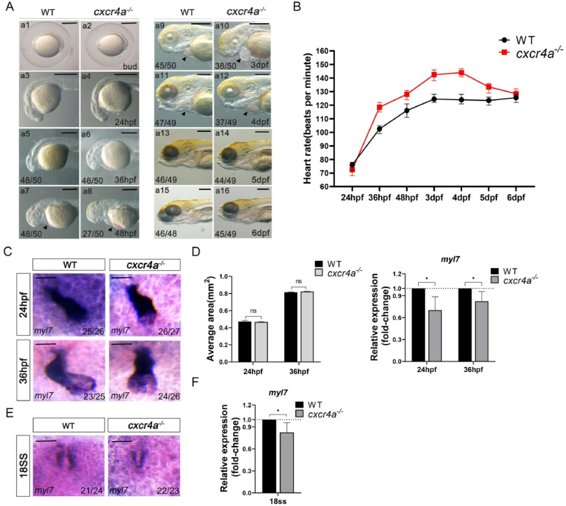
Fig. 1 Cardiac development is disturbed in the cxcr4a−/− embryos. (A) Comparing the wild-type (WT) and cxcr4a−/− embryos from bud stage to 6 dpf. The mutants display pericardial effusion and erythrocyte remaining from 2dpf to 4dpf (black arrow showed). However, the development of head and eyes are normal. (B) Knocking out cxcr4a increases the heart rate from 36hpf to 5dpf. The data represents average heart rate. Error bars indicate standard deviation of the mean. Unit: beats per minute. (C, D) The embryonic heart in cxcr4a−/− embryos is deformed, displaying defect heart jogging (C). But the heart size is normal (D). The expression of myl7 is examined using RT-qPCR and it is decreased in cxcr4a mutants at 24hpf and 36hpf when compared with WT embryos (D). (E, F) At 18 SS, the expression of myl7 is examined using in situ experiments and RT-qPCR, the expression of myl7 is decreased. Error bars indicate standard deviation of the mean. ns: not significant. Bar in image A, 50 μm; Bar in image C and E, 20 μm *P<0.05.