
|
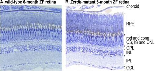
Fig. 2 Histological analysis of retina sections from wild-type and Zcrdh-mutant zebrafish Histologic sections from 6-month-old wild-type (A) and Zcrdh-mutant (B) zebrafish retinas. Sections were stained with 1% toluidine blue and 1% sodium borate and photographed with a 20× objective. Retina layers are identified to the right of (B). RPE, retinal pigment epithelium; rod and cone OS, IS, and ONL, rod and cone outer segments, inner segments, and outer nuclear layer (containing rod and cone photoreceptor nuclei); OPL, outer plexiform layer; INL, inner nuclear layer; IPL, inner plexiform layer; GCL, ganglion cell layer. Note the similar number of photoreceptor nuclei and similar thickness of retinal layers in wild-type and Zcrdh-mutant retinas.