
|
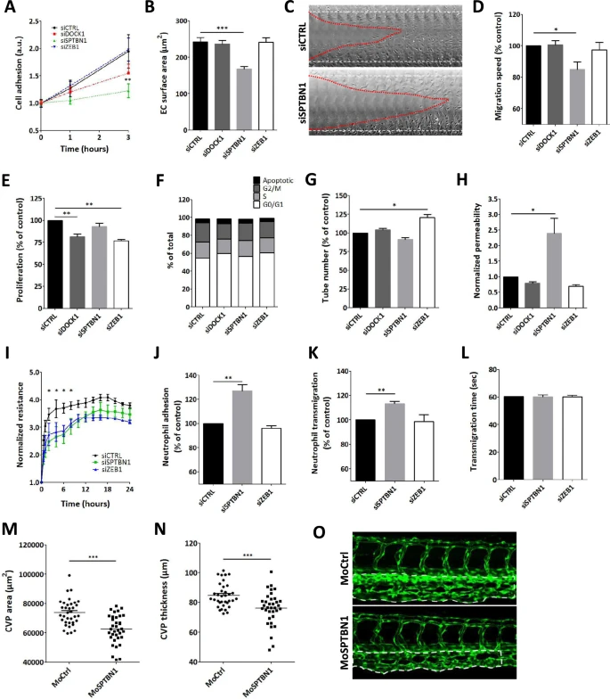
Fig. 3 Knockdown of dock1, sptbn1, and zeb1 reveal a range of functional effects. A Endothelial cell adhesion was significantly altered for sptbn1 knockdown, whereas dock1 knockdown showed an intermediate phenotype and siZEB1 was ineffective. B For sptbn1, the reduced cell adhesion is accompanied by defective cell spreading. C + D In addition, sptbn1 knockdown also reduced endothelial migration speed in a wound healing assay (C: example; D quantitative data). E Endothelial proliferation rate was impaired after dock1 and zeb1, but not sptbn1 knockdown; F cycle stage analysis did not reveal significant shifts in cell cycle stage. G Tube formation was slightly increased upon zeb1 knockdown, with a trend towards reduced tube formation in sptbn1 knockdown cells. H As a functional parameter, we assessed permeability for 150kD dextran in a Transwell system, revealing a significant increase in permeability upon knockdown of sptbn1, whereas dock1 or zeb1 silencing was ineffective. I In keeping, we observed decreased resistance of the endothelial monolayer upon sptbn11 but not zeb1 knockdown. J–L Furthermore, sptbn1 but not zeb1 knockdown also increased adhesion (J) and transmigration (K) of neutrophils under flow, while overall transmigration time (L) was unaffected. Based on these findings SNTBN1 was taken for in vivo validation in zebrafish. M, O Microinjection of zebrafish embryo’s with morpholino antisense oligonucleotides against sptbn1 led to a significant reduction in caudal vein plexus (CVP) development in zebrafish, as shown by the CVP area (M) and CVP thickness (N). Representative images of control and sptbn1 in zebrafish show de reduced CVP area and thickness (O, dotted area). Data are from three (A–G, I–L) or four (H) independent experiments or 35 zebrafish per group (M–O); error bars indicate SEM (A-C, E-N); *p ≤ 0.05; **p ≤ 0.01; ***p ≤ 0.005