
|
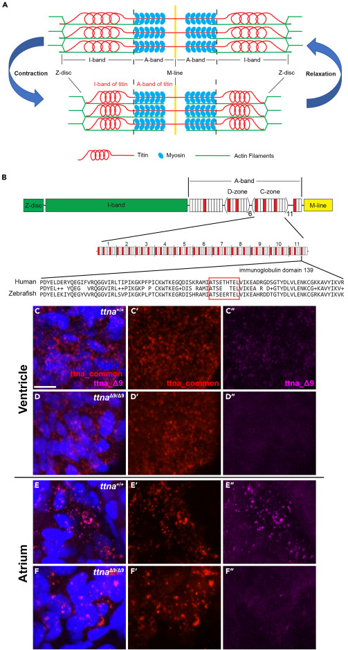
Fig. 1 ttnaΔ9 results in a 9 amino acid deletion in an immunoglobin-like domain corresponding to the human Ig-139 domain of titin A-band (A) Schematic of major components of the sarcomere. (B) Schematic of the full-length human titin protein and detailed layout of the C-zone in the A-band. Red, Ig domain. White, fibronectin type-III (FN3) domain. There are 11 repeats of Ig-FN3-FN3-Ig-FN3-FN3-FN3-Ig-FN3-FN3-FN3 pattern of domains in the C-zone. Amino acid sequence alignment between human Ig domain 139 (Ig-139) and its corresponding Ig domain (Ig-107) in the zebrafish ttna protein. Red box indicates the location of residues deleted in the TTN Δ9 and ttnaΔ9 mutation. (C–F″) Representative 3D projection images of in situ hybridization chain reaction with probes targeting ttna mRNA in either the 27 base pair deleted region (ttna_Δ9, magenta) or regions approximately 1,500 base pairs upstream and downstream of the deletion (ttna_common, red) in ventricular (C–D″) and atrial (E–F″) cardiomyocytes of wild-type (ttna+/+, n = 6) and ttnaΔ9/Δ9 (n = 6) zebrafish embryos at 48 h postfertilization (hpf). Optical section thickness: 29.8 μm. Scale bar: 10 μm. See also Figure S1 .