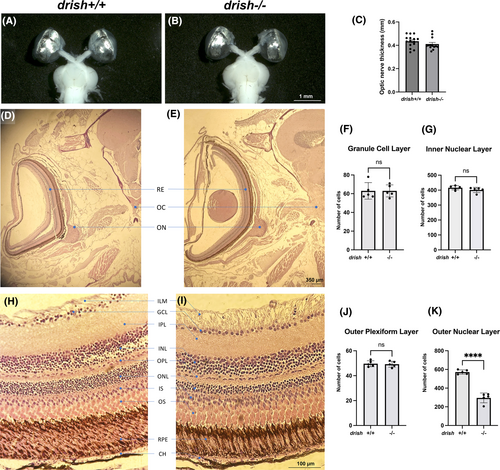
Fig. 10 Analysis of brain preparations and histological sections in drish mutant and wild-type sibling adults. (A–C) Brains and eyes were dissected from approximately 1-year old adults and the optic nerve diameter was measured. No significant difference between drish−/− (n = 13) and drish+/+ (n = 15) sibling adults was observed (Student's t-test, p > 0.05). (D, E) H&E stained coronal sections of drish+/+ (D) and drish−/− (E) head were observed to analyze morphology of optic nerve pathfinding and optic chiasma formation in approximately 2.5-year old adult zebrafish. No significant difference was observed (n = 5 fish for each genotype). (F–K) H&E stained coronal sections of drish+/+ (H) and drish−/− (I) adult retina were observed to analyze disruptions in different layers of cells within retina. No difference in the number of cells in the granule cell layer (F), inner nuclear layer (G) or outer plexiform layer (J) was observed. However, the number of cells in the outer nuclear layer of drish−/− retina was significantly reduced (Student's t-test, ****p < 0.001) compared to drish+/+ retina (K). BR, brain; CH, choroid; GCL, ganglion cell layer; ILM, inner limiting membrane; INL, inner nuclear layer; IPL, inner plexiform layer; IS and OS, inner and outer segments of photoreceptors; OC, optic chiasma; ON, optic nerve; ONL, outer nuclear layer; RE, retina; RPE, retinal pigment epithelium; PR, photoreceptor layer.
Image
Figure Caption
Acknowledgments
This image is the copyrighted work of the attributed author or publisher, and
ZFIN has permission only to display this image to its users.
Additional permissions should be obtained from the applicable author or publisher of the image.
Full text @ Dev. Dyn.

