
Fig. 7
- ID
- ZDB-IMAGE-240802-36
- Publication
- Labudina et al., 2024 - Cohesin composition and dosage independently affect early development in zebrafish
- All Figures
- Figures for Labudina et al., 2024
|
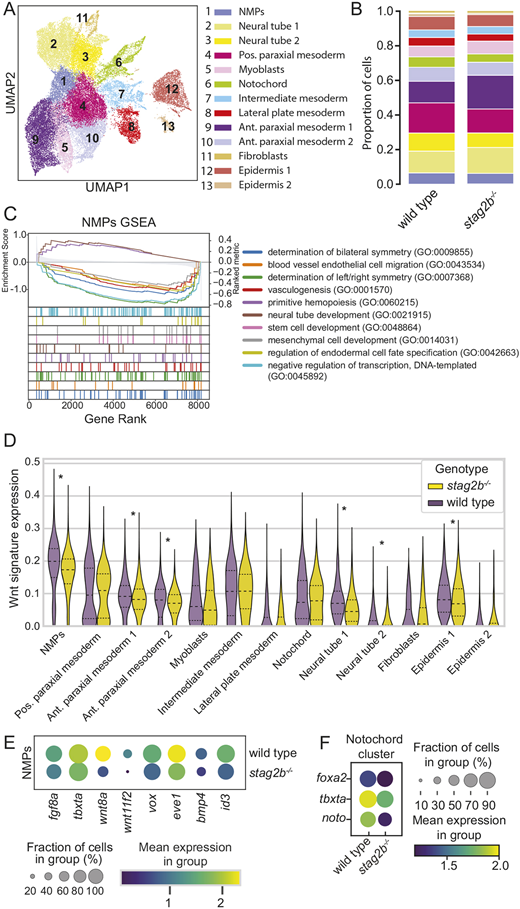
Fig. 7 Single-cell RNA-seq of tailbuds from embryos at the 16-somite stage shows disruption of Wnt signaling in stag2b−/− NMPs. (A) UMAP dimensional reduction of two integrated datasets of wild-type (15,298 cells) and stag2b−/− (21,278 cells) tailbud samples (total 36,576 cells) with clustering of the major cell types. (B) Stacked bar graph showing cell type proportions in wild type and stag2b−/−, color-coded according to the key in A. (C) Gene set enrichments for genes ranked by Z score for differential expression between wild-type and stag2b−/− NMP clusters. (D) Violin plot of Wnt gene expression signature (log-normalized) among different cell types in stag2b−/− (yellow) and wild-type (purple) embryos. Horizontal dashed lines represent 25th, 50th and 75th percentile. Wilcoxon rank-sum test with 5% FDR. *P<0.05. (E) Dot plot showing the mean expression of DEGs part of FGF, Wnt and BMP pathways in the NMP cluster in wild-type and stag2b−/− embryos. (F) Dot plot of DEGs in the notochord cluster.