
|
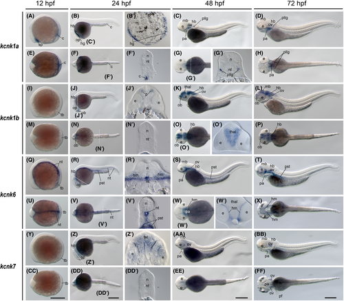
Fig. 4 The kcnk1a, kcnk1b, kcnk6, and kcnk7 gene (tandem of pore domains in a weak inward rectifying K+ [TWIK] channel) expression during zebrafish embryogenesis. Whole-mount in situ hybridization of zebrafish embryos at stages 12 h postfertilization (hpf) (A, E, I, M, Q, U, Y, and CC), 24 hpf (B, F, J, N, R, V, Z, and DD), 48 hpf (C, G, K, O, S, W, AA, EE), and 72 hpf (D, H, L, P, T, X, BB, and FF). The anterior is to the left in all the whole-mount images, and the dorsal is to the top in all transverse sections (B′, F′, G′, J′, N′, O′, R′, V′, W′, Z', and DD'). (A–D) Lateral view of gene expression of kcnk1a. (E–H) Dorsal view of gene expression of kcnk1a. (I–L) Lateral view of gene expression of kcnk1b. (M–P) Dorsal view of gene expression of kcnk1b. (Q–T) Lateral view of gene expression of kcnk6. (U–X) Dorsal view of gene expression of kcnk6. (Y–BB) Lateral view of gene expression of kcnk7. (CC–FF) Dorsal view of gene expression of kcnk7. The white dashed lines indicate the positions of sections left to the panel. The letters below or around the dashed lines correspond to the section panels. Scale bars were added on the bottom row of images; 250 μm for whole-mount images. c, cloaca; e, eye; hb, hindbrain; hg, hatching gland; hm, head mesenchymal region; mb, midbrain; n, neural tube; nt, notochord; ob, olfactory bulbs; op, optic vesicles; ov, otic vesicles; pa, pharyngeal arches; pf, pectoral fins; pllg, posterior lateral line ganglia; pst, pronephric proximal straight tubule; tb, tail bud; tel, telencephalon; thal, thalamus.