
|
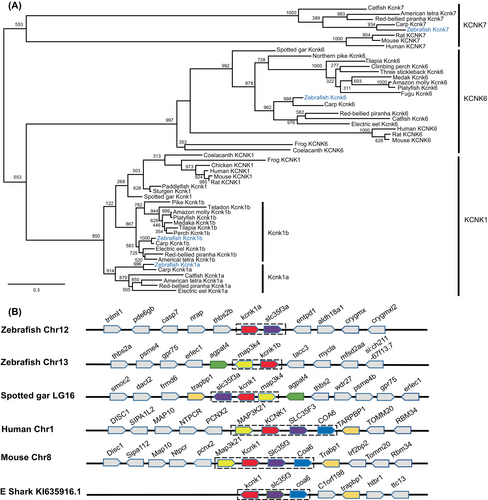
Fig. 3 The kcnk1 gene was duplicated in teleosts, including zebrafish. (A) Molecular phylogenetic tree generated using maximum likelihood analysis on vertebrate zebrafish tandem of pore domains in a weak inward rectifying K+ (TWIK) channel proteins as obtained with Jones–Taylor–Thornton model plus gamma distribution. Numbers at each node denote bootstrap values based on 1000 replicates. Branch lengths are proportional to expected replacements per site. All the zebrafish TWIK channels are highlighted in blue. KCNK1, KCNK6, and KCNK7 formed distinct clades. (B) The synteny of KCNK1 in five representative vertebrate species. The illustration of the genes and their sizes are not proportional to the length of the distances between genes. KCNK1 was highlighted in red. The conserved synteny (map3k4–kcnk1–slc35f3–coa6) was boxed with dashed lines.