IMAGE

Figure 1

ID
ZDB-IMAGE-240729-267
Source
Figures for Wei et al., 2024
Image
Figure Caption

Figure 1

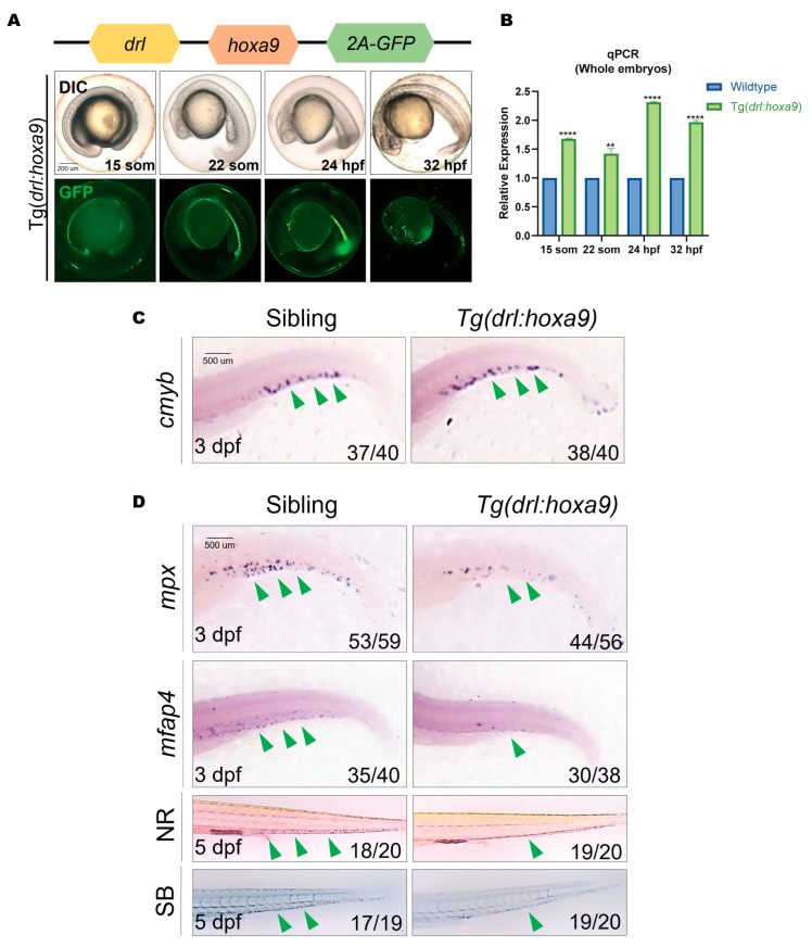
Myeloid development is arrested in Tg(drl:hoxa9) zebrafish embryos. (A) Schematic representation of the drl:hoxa9-2A-GFP reporter gene and the DIC and fluorescent images of Tg(drl:hoxa9) embryos. GFP is expressed in the lateral plate mesoderm and derived hematopoietic tissues at different stages. (B) RT-qPCR analysis of hoxa9 expression in Tg(drl:hoxa9) embryos compared to sibling embryos. Results are represented as mean ± SEM, t-test; ** p < 0.01, **** p< 0.0001. (C,D) WISH of cmyb (myeloid progenitor cells), mpx (granulocytes), and mfap4 (macrophages) in Tg(drl:hoxa9) and sibling embryos at 3 dpf. Neutral red (NR) and Sudan black (SB) staining of macrophages and neutrophils in embryos at 5 dpf. A green arrow indicates the staining signal. drl, draculin; WISH, whole mount in situ hybridization; hpf, hours post-fertilization; dpf, days post-fertilization.

Acknowledgments
This image is the copyrighted work of the attributed author or publisher, and ZFIN has permission only to display this image to its users. Additional permissions should be obtained from the applicable author or publisher of the image. Full text @ Int. J. Mol. Sci.