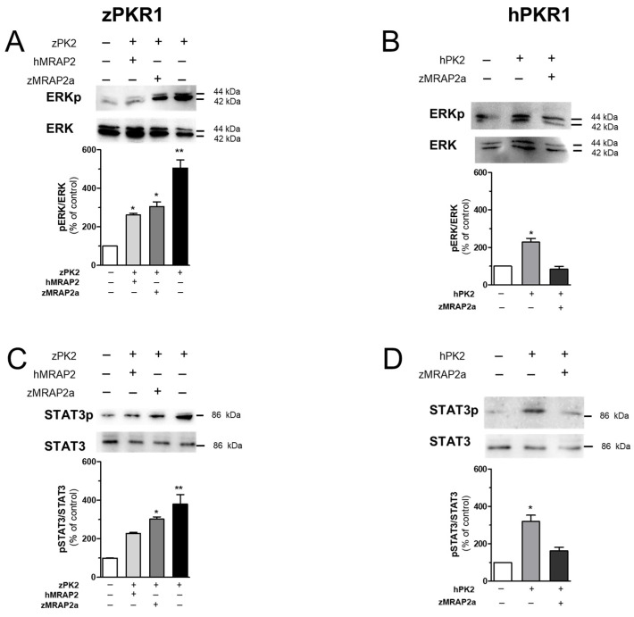
IMAGE

Figure 5

ID
ZDB-IMAGE-240729-192
Source
Figures for Fullone et al., 2024
Image
Figure Caption

Figure 5

Analysis of ERK and STAT3 activation in CHO cells. (A,B) Analysis of ERK1/2 phosphorylation in CHO cells transfected with zPKR1 or hPKR1. Densitometric plots show pERK1/2 and ERK1/2 protein levels 10 min after treatment with zPK2 or hPK2 (100 nM). The bar graphs show the pERK1/2/ERK1/2 ratio and the percentage increase compared to unstimulated cells (CTRL). (C,D) Densitometric plots show phospho-STAT3 (pSTAT3) and STAT3 protein levels in CHO cells expressing zPKR1 or hPKR1 after 1 h of treatment with zPK2 or hPK2 (100 nM). Data are presented as the ratio of pSTAT3 to total STAT3 protein and plotted as a percentage increase compared to CTRL. The bars show the mean values ± SEM of the three experimental conditions. For statistical analysis, a one-way ANOVA followed by Tukey’s test for multiple comparisons was used. * p < 0.05, ** p < 0.01 versus CTRL.

Acknowledgments
This image is the copyrighted work of the attributed author or publisher, and ZFIN has permission only to display this image to its users. Additional permissions should be obtained from the applicable author or publisher of the image. Full text @ Int. J. Mol. Sci.