
|
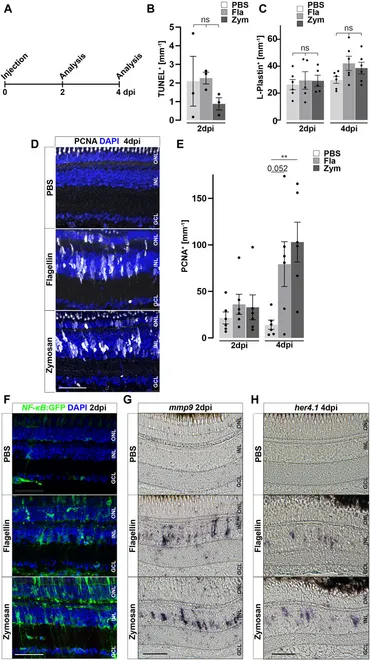
Fig. 6 Inflammatory stimuli trigger Müller glia reactivity. (A) Scheme of experimental outline. Wild type or NF-κB:GFP reporter animals were injected with flagellin or zymosan into the vitreous of the eye and analyzed at 2 and 4 days post injection (dpi). (B) Quantification of TUNEL+ nuclei in control and injected retinae at two dpi revealing no increase in cell death. See also Supplementary Figure S7 for counts of pyknotic nuclei. (C) Quantification of L-Plastin+ cells in control (PBS) and flagellin or zymosan injected retinae at two and four dpi display no significant increase in leukocytes due to the injection of factors. (D) Immunohistochemistry for proliferating cell nuclear antigen (PCNA) reveals proliferation in flagellin or zymosan injected but not in control animals at four dpi. Signal in the outer nuclear layer (ONL) appears to be a non-specific labeling of photoreceptor cells. (E) Quantification of PCNA+ nuclei in injected retinae at two and four dpi. (F) In comparison to PBS injected animals, injection of flagellin or zymosan results in strong activation of the NFκB:GFP reporter at two dpi. (G & H) In situ hybridization of mmp9 and her4.1 show expression of both genes in flagellin or zymosan injected, but not in PBS injected, eyes at two and four dpi, respectively. Retinal layering is indicated by dashed lines. Scale bar: 50 μm. Error bars indicate standard error; * * = p≤ 0.01 B: N = 3 Fish; C&E: N = 6 Fish; One-tailed ANOVA; ONL = outer nuclear layer; INL = inner nuclear layer; GCL = ganglion cell layer.