
|
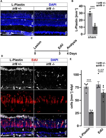
Fig. 5 Genetic reduction of microglia impairs retinal reactive proliferation in response to injury. (A) In contrast to heterozygous control siblings (irf8+/−), interferon regulatory factor 8 (irf8) homozygous myeloid-deficient mutant retinae (irf8−/−) display a decreased number of L-Plastin+ leukocytes in homeostasis. (B) Quantifications of L-Plastin+ cells in irf8−/− and control heterozygous siblings irf8+/−. (C) Scheme of experimental outline. irf8−/− and irf8+/− animals received an EdU pulse at 3 days post lesion (dpl) and were analyzed at 4 dpl. (D) During regeneration, the accumulation of L-Plastin+ cells at the outer nuclear layer (ONL) is impaired in irf8−/− but not in control irf8+/− siblings. Moreover, the number of EdU+ cells appears decreased in irf8−/− retinae. (E) Quantification of L-Plastin+ cells and EdU+ nuclei in irf8−/− and control irf8+/− animals at 4 dpl, showing reduced amounts of positive cells, respectively. Scale bar: 50 µm. Error bars indicate SEM; *** = p < 0.001; N = 5; two-tailed t-Test; INL = inner nuclear layer; GCL ganglion cell layer.