
|
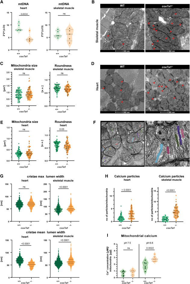
Fig. 2 Reduced mitochondrial performance in cox7a1−/− zebrafish (A) mtDNA content assessed by quantitative PCR on 5 cox7a1−/− or wild-type sibling female hearts and skeletal muscle samples. Shown are median and quartiles, as well individual data points. Unpaired t test. ns, non-significant. (B–G) Transmission electron microscopy of skeletal muscle and heart from adult fish (n = 3 males per genotype). Arrowheads, calcium precipitates. (B) and (D) Representative images of skeletal and cardiac muscle mitochondria. (C) and (E) Measurement of mitochondrial size and roundness in skeletal muscle or heart. Unpaired t test. (F) Zoomed-in views of mitochondria to show segmented cristae lumen. (G) Measurement of maximal cristae lumen width and average cristae lumen width. On average ∼250 cristae were analyzed per biological replicate. Mann-Whitney U test. (H) Number of calcium particles per mitochondria in heart and skeletal muscle. A minimum of 30 mitochondria per biological sample were measured from 3 to 5 different regions of the section. Biological samples are represented with different color tones. Unpaired t test. (I) Calcium levels in fresh adult skeletal muscle isolated mitochondria subjected to pH 7.5 (basal dissolved calcium levels) and pH 6.8 (solubilization of calcium precipitates) (n = 6 male biological replicates per genotype). Shown are individual measurements as well as median and quartiles two-way ANOVA, and Fisher’s LSD multiple comparison.
Reprinted from Developmental Cell, 59(14), García-Poyatos, C., Arora, P., Calvo, E., Marques, I.J., Kirschke, N., Galardi-Castilla, M., Lembke, C., Meer, M., Fernández-Montes, P., Ernst, A., Haberthür, D., Hlushchuk, R., Vázquez, J., Vermathen, P., Enríquez, J.A., Mercader, N., Cox7a1 controls skeletal muscle physiology and heart regeneration through complex IV dimerization, 1824-1841.e10, Copyright (2024) with permission from Elsevier. Full text @ Dev. Cell