
|
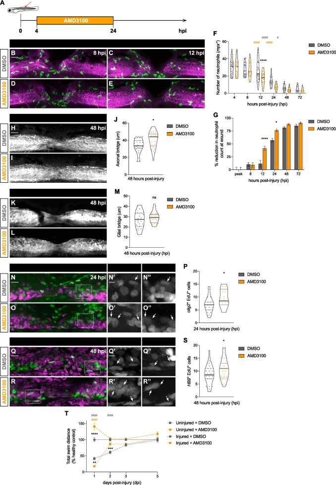
Fig. 3 AMD3100 fastens neutrophil clearance from the injured spinal cord and improves spinal cord regeneration. (A) Scheme represents the administration of AMD3100 3 dpf larvae, from 4 until 24 hpi. (B − E) Representative maximal projections display neutrophils (green) recruited to the injured spinal cord (magenta) at 8 and 12 hpi. Scale bar, 30 μm. (F) Quantification of neutrophil numbers at the injury site at 4, 8, 12, 24, 48 and 72 hpi (n = 40 (4, 8 and 12 hpi), 31 (24 hpi), 24 (48 hpi) and 19 (72 hpi), from three independent experiments). Two-way ANOVA followed by Tukey’s multiple comparisons test indicates a significant decrease between AMD3100- and DMSO-treated larvae at 12 hpi (p < 0.0001) (black asterisk) with no significant difference at any other timepoints. Significant differences were also observed within each treatment for DMSO-treated larvae (grey pounds) between 12 and 24 hpi (p < 0.0001) and 24 and 48 hpi (p = 0.0121), and AMD3100-treated larvae (orange pounds) between 8 and 12 hpi (p < 0.0001) and 12 and 24 hpi (p < 0.0001). (G) Quantification shows the percentage of reduction in neutrophil counts at the wound. Two-way ANOVA followed by Sidak’s multiple comparison test indicates a significant increase between AMD3100- and DMSO-treated larvae at 12 (p < 0.0001) and 24 hpi (p = 0.0134) and no significant difference at the peak and 8 hpi (p > 0.9999), 48 (p = 0.9282) and 72 hpi (p = 0.9870). (H, I) Representative maximal projections of the spinal cord, highlighting the axonal bridge. Scale bar, 30 μm. (J) Quantification of the axonal bridge thickness reveals a significant increase between AMD3100- and DMSO-treated injured larvae, analyzed via an unpaired two-tailed t-test (p = 0.0105) (n = 21–24, from two independent experiments). (K, L) Representative maximal projections showing the glial bridge. Scale bar, 30 μm. (M) Quantification of the glial bridge demonstrates no significant difference between AMD3100- and DMSO-treated larvae, analyzed using an unpaired two-tailed t-test (p = 0.2675) (n = 24–30, from two independent experiments). (N, O) Representative maximal projections of the spinal cord illustrate EdU incorporation in neural progenitors (olig2+ cells). Scale bar, 30 μm. Higher magnification panels (N’,O’) and (N’’, O’’) indicate regions outlined in rectangular boxes for the Edu and olig2 channels, respectively. Scale bar, 10 μm. (P) Quantification of Edu+ olig2+ cells in DMSO- and AMD3100- treated larvae (n = 19–20, from three independent experiments) shows a significant difference (p = 0.0213) via an unpaired two-tailed t-test. (Q, R) Representative maximal projections of the spinal cord exhibit EdU incorporation in motoneurons (HB9+ cells). Scale bar, 30 μm. Higher magnification panels (Q’, R’) and (Q’’,’R’) indicate regions outlined in rectangular boxes for the Edu and HB9 channels, respectively. Scale bar, 10 μm. (S) Quantification of Edu+ HB9+ cells in DMSO- and AMD3100- treated larvae (n = 19–20, from three independent experiments) shows a significant difference (p = 0.0428) via an unpaired two-tailed t-test. (T) Recovery of the swimming capacity, expressed as the total swim distance in relationship to the uninjured control (n = 40–71, from three independent experiments). Two-way ANOVA followed by Tukey’s multiple comparison test reveals significant difference between injured AMD3100- and DMSO-treated larvae (black asterisks) at 1 (p = 0.0099) and 2 dpi (p = 0.0002), between uninjured AMD3100- and DMSO-treated larvae (black asterisk) at 1 dpi (p < 0.0001), between injured and uninjured DMSO-treated larvae (grey pounds) at 1 (p < 0.0001) and 2 dpi (p < 0.0001); and between injured AMD3100- and uninjured DMSO-treated larvae (orange pounds) at 1 dpi (p < 0.0001).