
Fig. 2
- ID
- ZDB-IMAGE-240717-23
- Genes
- Publication
- Chen et al., 2024 - Epidermal growth factor-like domain 7 drives brain lymphatic endothelial cell development through integrin αvβ3
- All Figures
- Figures for Chen et al., 2024
|
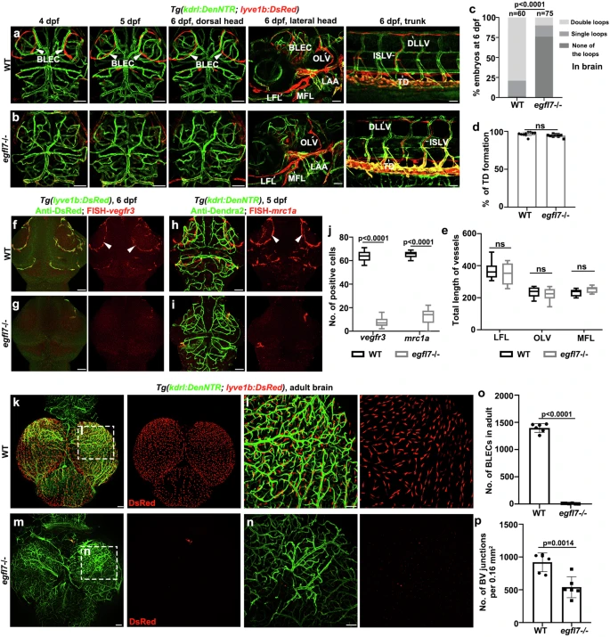
Fig. 2 egfl7 is essential for zebrafish BLEC formation.a Confocal images of the BLECs, facial lymphatics, and trunk lymphatics in Tg(kdrl: DenNTR;lyve1b: DsRed) transgenic lines from 4 dpf to 6 dpf in WT. b Loss of egfl7 prevents the formation of the BLECs from 4 dpf to 6 dpf, but the facial lymphatics of the lateral head and trunk lymphatics are normal in egfl7 mutant embryos at 6 dpf. c Percentage of embryos that have double lymphatic loops, single loops, and none of the loops in the brain (WT, n = 60 embryos, egfl7-/-, n = 75 embryos, χ2 test). d The statistics show the percentage of TD formation per 6 somites (n = 10 embryos, two-tailed unpaired t-test; ns, no significance. Data are represented as mean ± SD). e The statistics show the total lengths of LFLs, OLVs, and MFLs at 6 dpf (n = 8 embryos, 2way ANOVA multiple comparisons test; ns, no significance. Box plots show the five-number summary of a set of data: including the minimum score (shown at the end of the lower whisker), first (lower) quartile, median, third (upper) quartile, and maximum score (shown at the end of the upper whisker)). BLECs, brain lymphatic endothelial cells; LFL lateral facial lymphatic, MFL medial facial lymphatic, LAA lymphatic branchial arches, OLV otolithic lymphatic vessel, ISLV intersegmental lymphatic vessels, DLLV dorsal longitudinal lymphatic vessel, TD thoracic duct. f, g Double labeling of FISH-vegfr3 and anti-DsRed in Tg(lyve1b:DsRed) transgenic background at 6 dpf. Arrowheads point to the BLECs in the top layer of the brain. h, i Double labeling of FISH-mrc1a and anti-Dendra2 in Tg(kdrl:DenNTR) transgenic background at 5 dpf. j The statistics show the number of vegfr3+ and mrc1a + BLECs in WT and egfl7 mutant (n = 9 embryos, 2-way ANOVA multiple comparisons test. Box plots show the five-number summary of a set of data: including the minimum score (shown at the end of the lower whisker), first (lower) quartile, median, third (upper) quartile, and maximum score (shown at the end of the upper whisker). k–n Dissection and whole-mount images of BLECs and meningeal vascular in Tg(kdrl: DenNTR;lyve1b: DsRed) adult brain at 6 mpf (months post-fertilization) in WT (k, l) and egfl7 mutant (m, n). o, p The statistics show the number of lyve1b + BLECs in the adult brain (o, n = 6 brains; two-tailed unpaired t test) and the number of BV junctions per 0.16 mm2 of a brain (p, n = 6 brain areas; two-tailed unpaired t test). Data are represented as mean ± SD. Scale bar, 50 μm.