
Image
|
Figure Caption
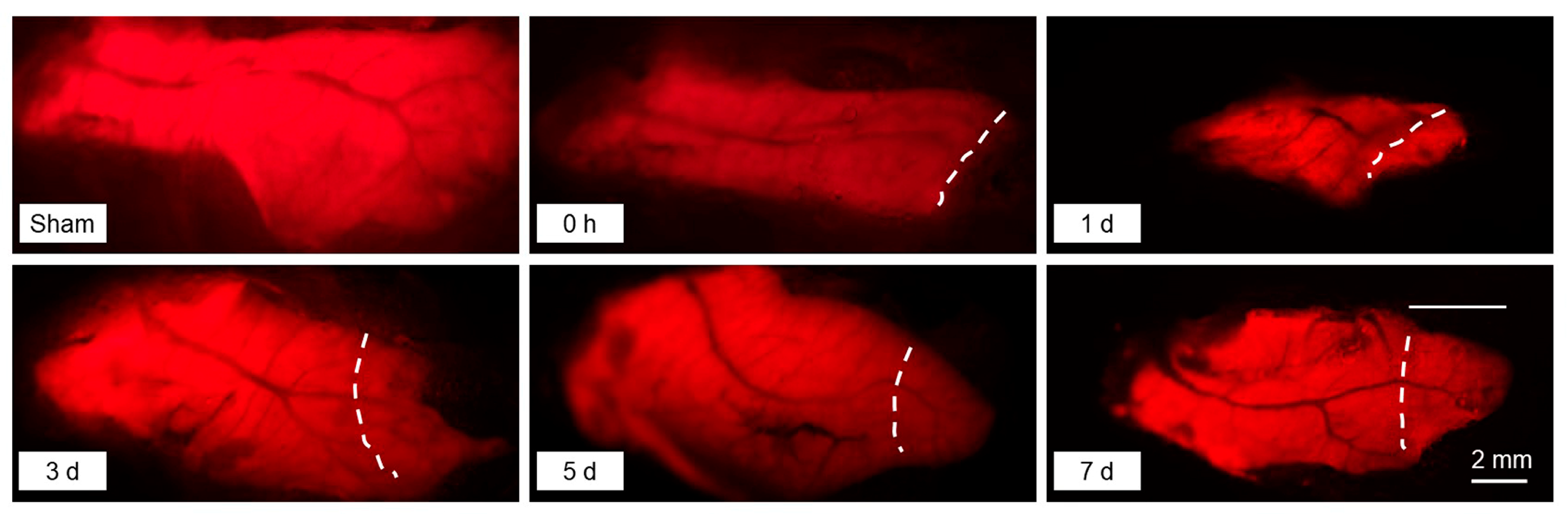
Fig. 1 Representative photos for the regenerating liver at the indicated times after the PHx surgery. The white dashed curves indicate the cutting edge resulting from the PHx surgery. The regions to the right of the curves are regenerated tissue after hepatectomy. The Tg(fabp10a:dsRed; ela3l:EGFP) transgenic fish line specifically expressing DsRed in the liver was used for the experiment.
Acknowledgments
This image is the copyrighted work of the attributed author or publisher, and
ZFIN has permission only to display this image to its users.
Additional permissions should be obtained from the applicable author or publisher of the image.
Full text @ Int. J. Mol. Sci.