
Fig. 5
- ID
- ZDB-IMAGE-240708-44
- Publication
- McKaige et al., 2024 - Mitochondrial abnormalities contribute to muscle weakness in a Dnajb6 deficient zebrafish model
- All Figures
- Figures for McKaige et al., 2024
|
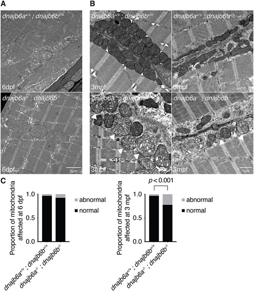
Fig. 5 Double dnajb6 mutants show mitochondrial abnormalities. (A) Electron microscopy of double mutant and wildtype muscle at 6 dpf shows no visible difference between double dnajb6 mutants and wildtype. (B) Ultrastructure of 3 mpf double mutant and wildtype muscle reveals mitochondria with abnormal cristae arrangement (arrow). (C) Quantification of mitochondrial abnormalities at 6 dpf and 3 mpf. At 6 dpf there was no sign of mitochondrial abnormalities in double mutants. However, at 3 mpf there was a significant increase in the proportion of abnormal mitochondria in double mutants compared to wildtype (P < 0.001, based on a binomial test). Analysis at both timepoints was completed blinded with two biological replicates and n = 4 per genotype.