
|
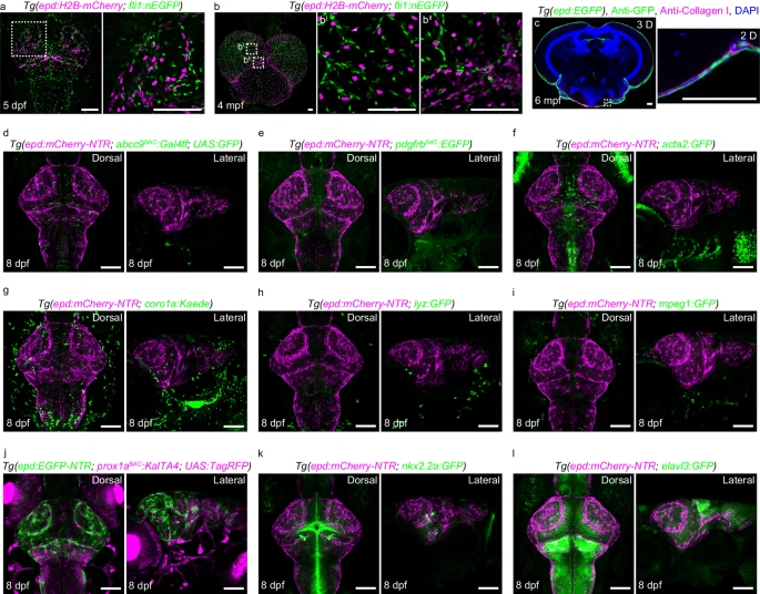
Fig. 2 The epd-positive cells do not belong to other known meningeal cell types.a, b Dorsal confocal images of epd:H2B-mCherry-positive nuclei and fli1:nEFP-positive nuclei in Tg(epd:H2B-mCherry; fli1:nEFP) brains at 5 dpf (a n = 20) and 4 mpf (b n = 8). c Confocal image of a cross-section of an adult brain showing collagen I-labeled fibroblasts at a different layer to epd-positive cells at 6 mpf. A 2D view of the enlarged area is shown. n = 8 adult brains. The experiment was repeated three times independently with similar results. d–l Confocal images showed epd-positive cells did not co-stain with abcc9BAC:Gal4ff; UAS:GFP-positive pericytes (d), pdgfrbBAC:EGFP-positive pericytes (e), acta2:GFP-positive smooth muscle cells (f), coro1a:Kaede-positive immune cells (g), lyz:GFP-positive neutrophils (h), mpeg1:GFP-positive macrophages (i), prox1aBAC:KalTA4; UAS:TagRFP-positive lymphatic endothelial cells and neuronal cells (j), nkx2.2a:GFP-positive neuronal and/or oligodendrocytes (k), and elavl3:GFP-positive neurons (l) at 8 dpf. Dorsal and lateral views of the larval brains were shown. n = 20 per experiment. Each experiment was repeated three times independently with similar results. The white dashed boxes outline the enlarged areas. Scale bars: 100 µm.