
|
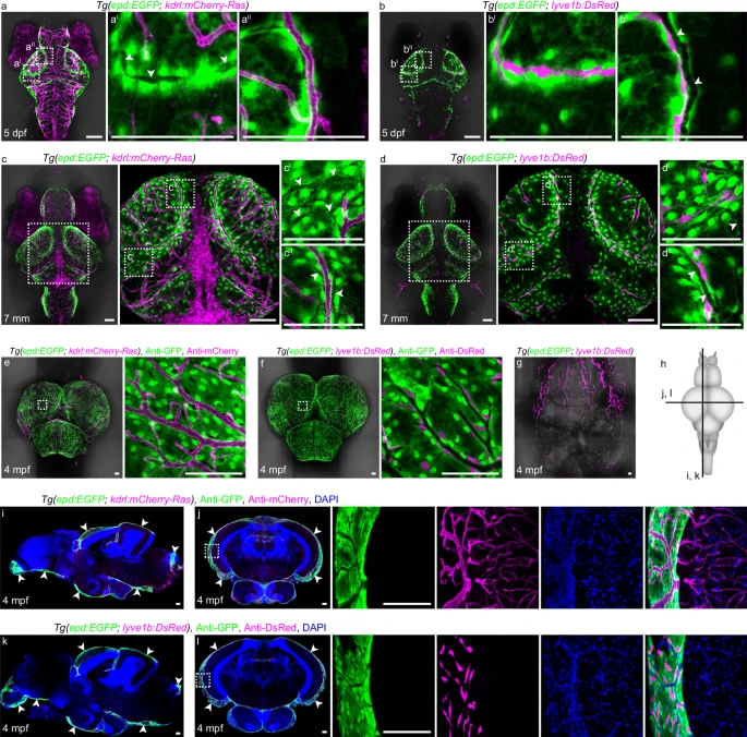
Fig. 1 The epd-positive cells in zebrafish leptomeninges consistently enwrap muLECs from larva to adult.a, c, e Dorsal confocal images of epd-positive cells and blood vessels on Tg(epd:EGFP; kdrl:mCherry-Ras) brains at 5 dpf (a n = 20), 7 mm (c n = 20) and 4 mpf (e n = 12). White arrowheads indicate the cavities of mural lymphatic endothelial cells (muLECs). b, d, f Dorsal confocal images of epd-positive cells and muLECs on Tg(epd:EGFP; lyve1b:DsRed) brains at 5 dpf (b n = 20), 7 mm (d n = 20) and 4 mpf (f n = 12). White arrowheads indicate the cavities of meningeal blood vessels. g A representative confocal image of the meningeal lymphatics in the dura mater beneath the skull at 4 mpf. n = 12. h Illustration of cross-sections (horizontal line) and sagittal sections (vertical line) of adult zebrafish brain in (i–l). i–l Confocal images of brain sections showing the locations of epd-positive cells in the leptomeninges at 4 mpf. White arrowheads indicate the distributions of epd-positive cells. n = 12 adult brains per experiment. Each experiment was repeated three times independently with similar results. The white dashed boxes outline the enlarged areas. Scale bars: 100 µm.