
|
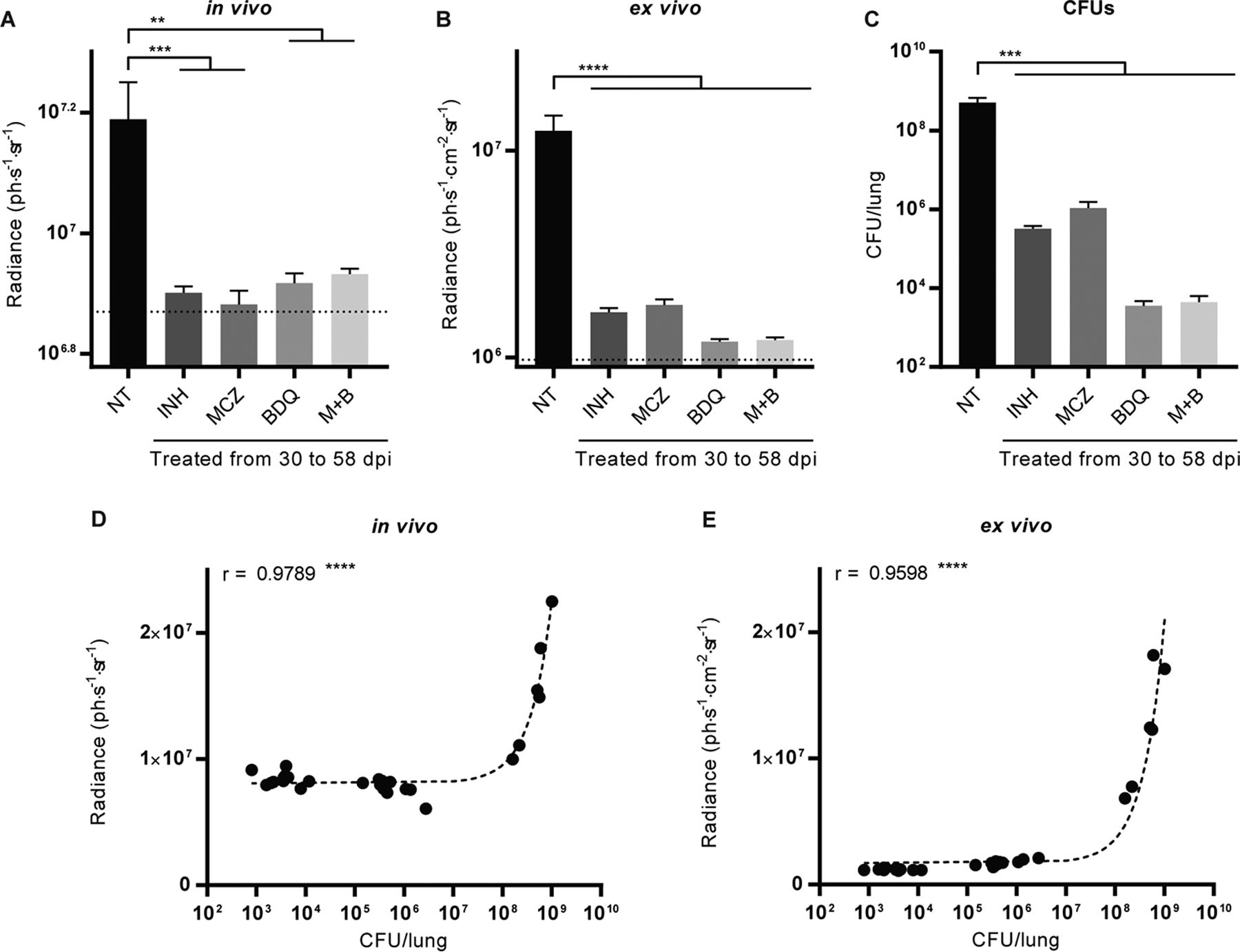
Fig. 5 Fluorescence measurements and CFU enumeration to assess antibiotic efficacy. (A, B) Fluorescence was measured in vivo and quantified in the thorax of mice (A) or postmortem on excised lungs (B). For the latter, the fluorescence intensity measurement is adjusted for the area of the lung on which the quantification was done. (C) Serial 10-fold dilutions of lung homogenates were plated, and the CFU were enumerated after 4 to 5 weeks of growth at 37°C. The dotted line indicates the average background fluorescence measured in uninfected mice (A) and lungs (B). Bars indicate the average ± SEM. Data are for 5 mice in each group. NT, no treatment; INH, isoniazid; MCZ, macozinone; BDQ, bedaquiline; M+B, a combination of macozinone and bedaquiline. **, P < 0.01; ***, P < 0.001; ****, P < 0.0001. (D, E) The number of CFU recovered at the end of the experiment from the lungs of untreated and treated mice correlates with the fluorescence measured and quantified in vivo (D) and ex vivo on excised lungs (E). Each point corresponds to a measurement for one mouse, r is the Pearson correlation coefficient, and dashed lines are linear regressions. ****, P < 0.0001.