
|
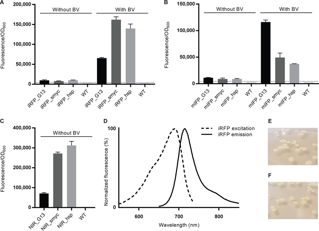
Fig. 1 M. smegmatis reporter strains emit NIR fluorescence. (A to C) Fluorescence intensity measured at 713 nm for M. smegmatis expressing iRFP (A) or mIFP (B) and cultured overnight with or without BV or for NIR fluorescent M. smegmatis reporters without BV supplementation (C). The fluorescence intensity was measured with a microplate reader and normalized according to the cell density for each well. The dotted lines indicate the background autofluorescence recorded in WT bacteria under the same conditions. Data represent the averages ± SEM for 4 independent clones per group. (D) Fluorescence excitation and emission spectra of the M. smegmatis NIR_smyc reporter. (E, F) Photographs of colonies of WT M. smegmatis (E) and M. smegmatis NIR_smyc reporter bacteria (F), with the latter displaying a faint green color.