
Fig. 3
- ID
- ZDB-IMAGE-240625-48
- Publication
- Jędrychowska et al., 2024 - Mutant analysis of Kcng4b reveals how the different functional states of the voltage-gated potassium channel regulate ear development
- All Figures
- Figures for Jędrychowska et al., 2024
|
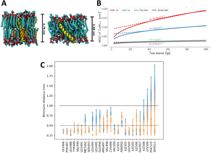
Fig. 3 Characterization of S7 domain in the Kcng4b-C2 mutants. A - Representations of isolated helix systems of Kcng4b-C2; S7 in a thicker, more stiff liquid ordered (Lo) phase (left), S7 in a thinner, looser liquid disordered (Ld) phase (right). B - Plots of mean square displacement (MSD) against time interval τ. Helices described as first and second refer to the S7s from two different Kcng4b/Kv6.4 monomers in the tetramer. Here, MSD measures displacement of the center of mass of the helix in membrane plane within the time interval τ. The slope of the MSD at long times is related to the diffusion coefficient D, equivalent to one obtained by standard experimental methods, e.g., single-molecule tracking. C - Distributions of minimum distances between the residues of S7 helices and the remaining membrane-bound part of the channel. Distributions for each of the helices are shown separately in blue and orange.
Reprinted from Developmental Biology, 513, Jędrychowska, J., Vardanyan, V., Wieczor, M., Marciniak, A., Czub, J., Amini, R., Jain, R., Shen, H., Choi, H., Kuznicki, J., Korzh, V., Mutant analysis of Kcng4b reveals how the different functional states of the voltage-gated potassium channel regulate ear development, 50-62, Copyright (2024) with permission from Elsevier. Full text @ Dev. Biol.