IMAGE

Fig. 6

ID
ZDB-IMAGE-240621-84
Source
Figures for Wang et al., 2024
Image
Figure Caption

Fig. 6

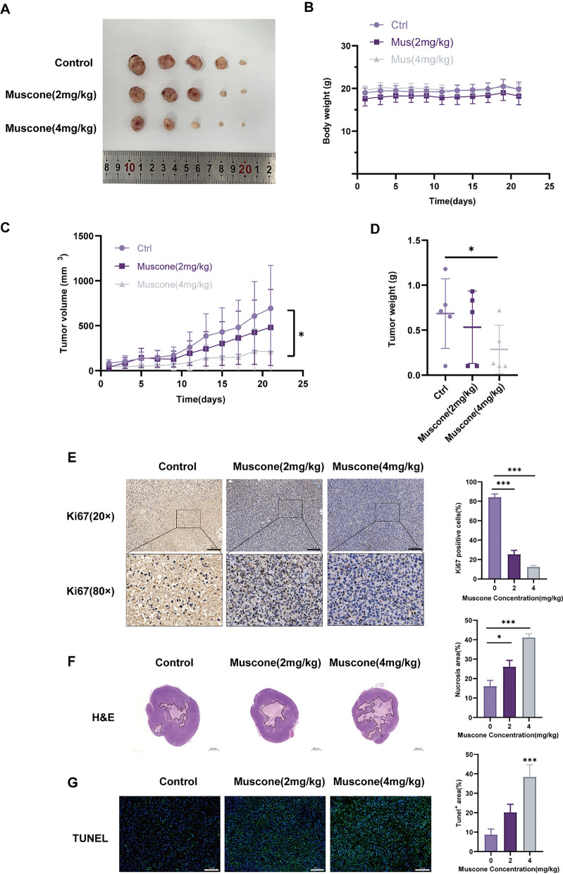
Muscone abolishes tumor development in a mouse breast cancer (BC) xenograft model. A Image of MDA-MB-231 tumors from different groups. B Body weights were recorded once every 2 days for 21 d. C Tumor volumes were computed once every 2 days. D Dissected tumor weights were accurately documented. E Representative immunohistochemical (IHC) Ki67 staining in MDA-MB-231 xenograft tumors; Scale bar, 200 µm. F Tumor tissue H&E staining; Scale bar, 2000 µm. G Tumor tissue TUNEL staining; Scale bar, 100 µm. Representative staining images and quantification. Data provided as mean ± SEM. n = 5 mice per group. *P < 0.05, **P < 0.01, and ***P < 0.001

Acknowledgments
This image is the copyrighted work of the attributed author or publisher, and ZFIN has permission only to display this image to its users. Additional permissions should be obtained from the applicable author or publisher of the image. Full text @ Cancer Cell Int.